


Odpowiedź bezpośrednia
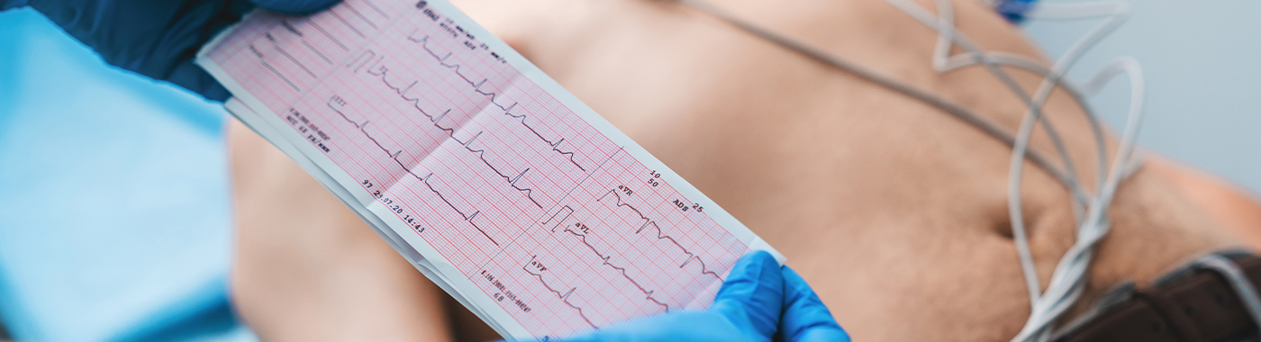
An electrocardiogram (ECG, also called EKG or "heart tracing") records the heart's electrical activity through electrodes placed on the chest and limbs. The ECG detects rhythm disturbances, conduction block, and many heart attacks. A normal ECG does not exclude coronary artery disease, and the tracing must be interpreted alongside symptoms.
Kluczowe dania na wynos
- Szybko i nieinwazyjnie: a standard 12-lead ECG takes a few minutes and involves no radiation and no injections.
- Rhythm and electrical events are its strength: arrhythmias, heart block and ST-segment changes of an evolving heart attack.
- A normal ECG is reassuring but not conclusive: many people with significant coronary narrowing have a normal resting tracing.
- To jest migawka: intermittent palpitations may need Holter or longer-term rhythm monitoring.
- Interpretacja wymaga kontekstu: the same tracing can mean different things in a 20-year-old athlete and a 65-year-old with diabetes.
W skrócie
| Cecha | Szczegół |
|---|---|
| Co mierzy | Electrical impulses generated by the heart, recorded from the body surface |
| Typ | Non-invasive, no radiation, no contrast |
| Czas nagrywania | Usually about 10 seconds of tracing; total visit commonly 5-10 minutes including electrode placement |
| Przygotowanie | Usually none; no fasting required for a standard resting ECG |
| Niewygoda | Minimal; adhesive pads may tug on removal, and chest hair may need trimming |
| Główne mocne strony | Heart rhythm, rate, conduction delays, acute ST-segment changes, some chamber enlargement patterns, some electrolyte and drug effects |
| Główne martwe pola | Coronary anatomy, valve function, pumping strength, intermittent arrhythmias |
| Kto to zgłasza | A physician or cardiologist, read together with your symptoms and history |
Znany także jako
- ECG (Indian and British usage) and EKG (from the German elektrokardiogram, common in the US)
- 12-lead ECG, resting ECG
- "Heart tracing", "heart graph", "ECG test"
- Commonly confused in conversation with the "echo test" (echocardiogram), which is a different examination using ultrasound
What an electrocardiogram is
The heart beats because of an electrical signal that starts in the sinoatrial (SA) node, spreads across the atria, pauses briefly at the atrioventricular (AV) node, and then travels down the conduction system to the ventricles. These currents reach the skin surface, where electrodes can pick them up.
A standard 12-lead ECG uses ten sticky electrodes, six on the chest and four on the limbs, to create twelve different electrical "views" of the heart. The machine prints a tracing with recognisable parts:
- Fala P: electrical activation of the atria.
- Zespół QRS: electrical activation of the ventricles.
- Fala T: electrical recovery (repolarisation) of the ventricles.
- Intervals (PR, QRS duration, QT): how long each step takes, which is where conduction delays and some drug effects show up.
The ECG records electrical behaviour only. The electrocardiogram does not image heart muscle, valves or arteries.
Why an electrocardiogram is done
- Chest pain or chest discomfort: to look for changes suggesting an acute coronary event, ideally within minutes of arrival in an emergency setting.
- Palpitations, fast or slow pulse: to characterise a rhythm if it is present during recording.
- Breathlessness, blackouts or near-fainting: to look for conduction block, pre-excitation patterns, long QT or evidence of strain.
- Znana choroba serca: to track conduction, rhythm and repolarisation over time.
- Medication monitoring: several cardiac drugs, some antipsychotics, some antibiotics and some antiemetics affect the QT interval or conduction.
- Electrolyte and kidney problems: marked potassium or calcium disturbance can change the tracing, though the ECG is not a substitute for a blood test.
- Before anaesthesia or surgery: when age, symptoms or existing heart disease make it relevant.
- Baseline before certain treatments: for example before starting drugs known to prolong the QT interval.
Kto powinien wykonać to badanie
An ECG is appropriate when a doctor has a specific question about your heart's electrical activity. That typically includes people who:
- have chest pain, pressure, or pain radiating to the jaw, back or arm, urgently
- have palpitations, an irregular pulse, or documented fast/slow heart rate
- have fainted, especially during exertion, or have a family history of sudden cardiac death in a young relative
- have unexplained breathlessness or reduced exercise tolerance
- live with high blood pressure, diabetes, chronic kidney disease, or established coronary or valvular disease, including rheumatic heart disease
- are on medicines that can affect heart rhythm or the QT interval, as advised by the prescriber
- are being assessed before surgery, or as part of a structured pre-participation sports assessment where local protocol requires it
A resting ECG in a young, symptom-free person with no risk factors often adds little and can raise questions about harmless variants. Whether a screening ECG is useful is a decision for your doctor, not a routine add-on. Results vary depending on individual clinical circumstances.
What an electrocardiogram cannot detect or exclude
This is the single most misunderstood part of the test.
- Badanie to nie uwidacznia tętnic wieńcowych. A normal resting ECG does not exclude coronary artery disease, and does not tell you whether an artery is narrowed.
- It does not measure pumping strength. Ejection fraction comes from an echocardiogram or cardiac MRI, not an ECG.
- It does not assess valves. Valve narrowing or leakage, including rheumatic valve disease, needs echocardiography. An ECG may show secondary chamber patterns, but cannot grade a valve.
- It cannot reliably exclude a heart attack. An early or non-ST-elevation myocardial infarction can have a normal or near-normal initial tracing. Serial ECGs plus high-sensitivity troponin blood tests are used, because a single tracing is not enough.
- It usually misses intermittent arrhythmias. A 10-second recording captures the rhythm only at that moment. If palpitations come and go, a normal ECG does not exclude an arrhythmia; Holter, extended patch or event monitoring may be needed.
- It does not diagnose blood clots in the lung directly. Pulmonary embolism may produce suggestive patterns, but diagnosis relies on imaging such as CT pulmonary angiography.
- Chamber enlargement patterns are indirect. ECG criteria for left ventricular hypertrophy are neither sensitive nor specific; echocardiography measures it.
- Some structural and genetic conditions can have a normal ECG, particularly early on.
"Not identified" is not the same as "excluded". If your chest pain, breathlessness, palpitations or fainting continue despite a normal ECG, go back to your doctor, do not treat the normal report as a closed file.
Powiązane, ale różne badania
| Testowanie | Pytanie, na które odpowiada | Na co nie odpowiada |
|---|---|---|
| Spoczynkowe EKG 12-odprowadzeniowe | What is the heart's electrical activity right now? | Coronary anatomy, pumping function, valve disease, intermittent rhythms |
| Holter / extended rhythm monitor | What is the rhythm over 24 hours to several days or weeks, and does it match symptoms? | Coronary narrowing; structural detail |
| Exercise (stress) ECG / TMT | Czy podczas wysiłku pojawiają się zmiany w EKG lub inne objawy? | Cannot map coronary anatomy; false positives and negatives are common, and results depend on pre-test probability |
| Echocardiogram ("echo test") | How well do the chambers pump, and how do the valves and pericardium look? | Does not visualise coronary arteries; a normal resting echo does not exclude coronary artery disease |
| Stress echo / nuclear perfusion imaging | Is there evidence of reduced blood supply to a territory of muscle under stress? | Does not give anatomical detail of the artery |
| Koronarografia TK | Are the coronary arteries narrowed or calcified, anatomically? | Uses radiation and iodinated contrast; does not directly measure rhythm over time |
| Inwazyjna koronarografia | Anatomy with the option of treating a blockage in the same sitting | Invasive, with procedural risks |
| Troponin blood test | Czy mięsień sercowy uległ uszkodzeniu? | Does not show rhythm or the site of injury |
| Badanie elektrofizjologiczne | Where exactly does an arrhythmia arise, and can it be ablated? | Invasive; reserved for selected rhythm problems |
No single test is universally "better". Which test comes first depends on the problem: ongoing chest pain in an emergency department starts with an ECG and troponin; recurrent palpitations usually needs rhythm monitoring; a murmur or suspected valve disease points to echocardiography; stable chest pain in someone at intermediate risk often leads to CT coronary angiography or functional imaging, depending on availability and the treating team's judgement.
Jak przygotować
Preparation for a resting ECG is minimal, and specific instructions are operational and set by the performing unit. Your unit's written instructions take precedence over anything you read online.
- Odzież: wear something that opens or lifts easily at the chest; you may be asked to change into a gown.
- Skóra: avoid oil, moisturiser, talc or lotion on the chest, arms and legs on the day, as these reduce electrode contact. Chest hair may need clipping at the electrode sites.
- Post: not required for a standard resting ECG. If the ECG is part of a stress test or a wider package, follow the specific protocol for that test.
- Leki: bring your current medicine list. Do not stop, start or alter any prescribed medicine for the test, including beta blockers, antiarrhythmics or thyroid medication, unless the doctor who prescribed it tells you to.
- Środki pobudzające: for a resting ECG, caffeine and nicotine are not an absolute barrier, but they can raise the heart rate and add ectopic beats. If the test is being done specifically to evaluate palpitations or for a stress test, follow the unit's guidance.
- Powiedz personelowi if you have a pacemaker or implanted defibrillator, previous heart attack or bypass surgery, chest wounds or dressings, or if you have had a similar test before, a previous tracing is valuable for comparison.
Ciąża
An ECG involves no radiation and is used in pregnancy when clinically indicated. Position may be adjusted, lying flat on the back in later pregnancy can cause light-headedness, so a left-tilted or semi-upright position is often used. Some ECG changes, including shifts in axis and minor T-wave changes, occur normally in pregnancy and need interpretation by a clinician aware of the pregnancy.
Dzieci
Paediatric ECGs use smaller electrodes and, importantly, age-specific normal values, adult criteria misread children's tracings. Explaining the stickers beforehand helps; movement and crying commonly cause artefact, so repeat recording may be needed.
Osoby starsze i osoby o ograniczonej sprawności ruchowej
Fragile or thin skin may react to adhesive, and tremor or Parkinson's disease can create baseline artefact. Tell the technician if lying flat is difficult; a semi-recumbent recording can be annotated.
People with pacemakers or implanted devices
An ECG is safe with pacemakers and implantable defibrillators and is often used precisely to check device behaviour. Always mention the device, as the tracing is interpreted differently. A device-specific interrogation by the cardiology team gives information the surface ECG cannot.
Cukrzyca, przewlekła choroba nerek i niewydolność serca
No special preparation is needed for a resting ECG, and no fluid or fasting restriction applies. If your ECG is part of a stress test, the fasting and medication instructions for that test can affect people on insulin or other glucose-lowering drugs, people on fluid restriction, and people with advanced kidney disease or heart failure, ask the requesting doctor for individualised instructions before you fast or skip a dose.
What happens during an electrocardiogram
- Pozycjonowanie: you lie on a couch and expose the chest, wrists and ankles. Privacy and a chaperone can be requested.
- Umieszczenie elektrod: ten adhesive electrodes are placed at defined anatomical positions. Accurate placement matters; misplaced leads are a frequent cause of odd-looking tracings.
- Nagranie: you lie still, breathe normally and avoid talking for a few seconds while the machine records. Shivering, coughing or movement can blur the tracing, and the technician may repeat it.
- Następnie: electrodes are peeled off. Brief skin redness or slight stickiness is common. Normal activity, eating and driving can resume immediately, unless the reason for the test itself imposes restrictions.
Jak długo trwa proces
The actual electrical recording is usually about 10 seconds. Electrode placement, positioning and undressing/dressing mean the appointment commonly lasts 5-10 minutes, and longer if repeat tracings are needed, if chest hair must be clipped, or in children. In an emergency department with chest pain, the aim is to have a tracing taken and reviewed within minutes of arrival. In an urgent setting a doctor reviews the tracing immediately; for routine outpatient tests, reporting time depends on the unit's workflow.
Zrozumienie raportu EKG
An ECG report usually has three parts: the printed tracing, a set of machine-generated measurements, and a human interpretation.
- Tętno: reported in beats per minute. Rates below or above the conventional adult range are not automatically abnormal, trained athletes often have slow resting rates, and anxiety, fever, pain, anaemia or dehydration commonly raise the rate.
- Rytm: "sinus rhythm" means the impulse is starting in the normal place. Atrial fibrillation, atrial flutter, supraventricular tachycardia and ventricular arrhythmias are described by name.
- Interwały: PR interval (AV conduction), QRS duration (ventricular conduction, prolonged in bundle branch block), and QT/QTc (repolarisation, relevant to certain drugs and inherited conditions).
- Axis and voltages: direction and size of the electrical forces. Voltage criteria for hypertrophy are unreliable on their own and are influenced by body build, chest wall thickness and lead placement.
- ST segment and T waves: ST elevation or depression and T-wave inversion may indicate ischaemia or injury, but also occur with pericarditis, myocarditis, electrolyte disturbance, bundle branch block, early repolarisation variants and left ventricular hypertrophy.
- Q waves: may indicate an old, healed heart attack, or may be positional and harmless.
The machine's automated statement at the top of the printout, phrases such as "abnormal ECG", "possible old infarct", "non-specific T-wave abnormality" or "consider left ventricular hypertrophy", is a computer suggestion, not a diagnosis. Automated readings are frequently overcalled and frequently corrected by the reporting doctor. Do not act on the printed statement before a clinician has interpreted the tracing in the context of your symptoms, examination, risk factors and previous ECGs. Comparison with an older tracing often changes the meaning entirely: a stable finding is far less worrying than a new one.
Kiedy wyniki EKG mogą być mylące
Wyniki, które mogą wydawać się fałszywie nieprawidłowe
- Lead misplacement or swapped limb leads can mimic old infarction, axis shift or even suggest dextrocardia.
- Movement, shivering, tremor or loose electrodes can imitate arrhythmia or produce a wandering baseline.
- Zakłócenia elektryczne from nearby equipment can overlay artefact that resembles flutter or fibrillation.
- Normal variants such as early repolarisation, sinus arrhythmia with breathing, and athlete-related changes are frequently flagged as "abnormal" by software.
- Body habitus, breast tissue, obesity, lung hyperinflation or oedema alter voltages and ST-T appearances.
- Bundle branch block, pacing and pre-excitation distort ST segments, making ischaemia assessment unreliable by surface ECG alone.
- Electrolyte and metabolic disturbance: potassium, calcium and magnesium abnormalities, and some drugs can produce ST-T and QT changes unrelated to coronary disease.
Findings that can look falsely normal or reassuring
- Early heart attack: the first tracing can be normal. Repeat ECGs and troponin testing are essential if symptoms suggest an acute coronary syndrome.
- Posterior or lateral territory infarction can be subtle on standard 12 leads.
- Przerywane arytmie vanish between episodes, so the resting tracing looks entirely normal.
- Stable coronary disease frequently produces a normal resting ECG even with significant narrowing.
- Baseline conduction abnormality or a paced rhythm może maskować nowe zmiany niedokrwienne.
Because of this, a doctor weighs the ECG against your story. Persistent or worsening symptoms outrank a normal tracing.
Ryzyko i bezpieczeństwo
Spoczynkowe EKG 12-odprowadzeniowe
A resting ECG records electrical signals; it does not deliver current into the body, uses no radiation and needs no injection. The practical issues are minor: transient skin redness or itching from adhesive, mild discomfort when pads are removed, discomfort from clipping chest hair, and rarely a local skin reaction. The main clinical risk is not physical but interpretive, a misread or over-interpreted tracing can lead to unnecessary anxiety and further testing, or to false reassurance.
Exercise (stress) ECG
A stress ECG carries a different and higher risk profile, because it deliberately increases cardiac workload. Uncommon but recognised events include chest pain, arrhythmia, marked blood pressure change, fainting and, rarely, serious cardiac events. It is performed with medical supervision and resuscitation facilities available, and it is not suitable for everyone, for example in suspected acute coronary syndrome, severe aortic stenosis or uncontrolled arrhythmia. Safety statements for a resting ECG do not transfer to a stress test.
Holter and ambulatory monitoring
Risks are limited to skin irritation from prolonged electrode contact and the inconvenience of wearing the device. Diagnostic yield depends on how often symptoms occur during the monitoring window.
czerwone flagi
W nagłym wypadku: zadzwoń na pogotowie lub udaj się do najbliższego oddziału ratunkowego
- Chest pain, pressure, heaviness or burning lasting more than a few minutes, or spreading to the arm, jaw, neck or back, especially with sweating, nausea or breathlessness
- Nagła, silna duszność lub duszność w spoczynku
- Fainting or collapse, particularly during exertion
- Palpitations with chest pain, fainting, severe breathlessness or confusion
- A very fast or very slow pulse with feeling unwell, cold, clammy or faint
- Sudden weakness or numbness of face, arm or leg, or difficulty speaking
- Do not wait for an outpatient ECG appointment in any of these situations.
Tego samego dnia: skontaktuj się ze swoim lekarzem już dziś
- New or worsening palpitations without the features above
- Breathlessness on mild exertion that is new for you
- Light-headedness or near-fainting on standing or on exertion
- New ankle swelling with breathlessness
- An ECG report handed to you with wording such as ST elevation, complete heart block, marked QT prolongation or ventricular tachycardia, if no doctor has yet reviewed it with you
- Symptoms starting soon after a new medicine known to affect heart rhythm, speak to the prescriber before making any change
Rutynowe spotkanie
- Occasional brief skipped-beat sensations with no other symptoms
- A machine statement such as "non-specific T-wave abnormality", "sinus bradycardia" in a fit person, or "possible left ventricular hypertrophy", so a clinician can compare with previous tracings
- Follow-up of known hypertension, diabetes, kidney disease or valve disease
- Family history of early heart disease or sudden cardiac death that you want assessed
Sytuacje specjalne
Ciąża
Palpitations and breathlessness are common in pregnancy, and an ECG is a reasonable, radiation-free first step. Physiological changes shift the tracing, so pregnancy-aware interpretation matters. Rheumatic valve disease presenting for the first time in pregnancy is relevant in India, and usually needs echocardiography rather than ECG alone.
Dzieci i młodzież
Paediatric tracings must be read against age-specific values. Investigation is warranted for fainting during exercise, palpitations with collapse, or a family history of sudden death in a young relative. Congenital heart disease assessment relies on echocardiography, with the ECG as a supporting test.
Starsi dorośli
Atrial fibrillation and conduction disease become more common with age, and heart attacks more often present without classic chest pain, with breathlessness, confusion or fatigue instead. Older tracings often show several coexisting abnormalities, so comparison with previous ECGs is especially useful.
Cukrzyca
Coronary disease can be more extensive and less symptomatic in people with long-standing diabetes, so a normal ECG should not be taken as clearance. Discuss any exertional symptom, even if mild, with your doctor.
Przewlekła choroba nerek i dializa
Potassium and calcium shifts around dialysis can change the tracing, and left ventricular hypertrophy is common. ECG changes are suggestive but do not replace biochemical testing for electrolyte disturbance.
Niewydolność serca
The ECG helps identify rhythm problems and conduction delay such as left bundle branch block, which influences device and treatment decisions. It does not measure ejection fraction; echocardiography and natriuretic peptide testing serve that purpose.
Sportowcy
Training produces recognised ECG changes, including a slow rate and increased voltages, which are not disease. Distinguishing these from cardiomyopathy or channelopathy needs athlete-specific criteria and often additional imaging.
The electrocardiogram in the Indian context
- Dostępność: ECG machines are among the most widely distributed diagnostic devices in India, present in district hospitals, small clinics, diagnostic labs and many primary health centres. This makes the ECG a practical first electrical assessment in both urban and rural settings.
- Language and naming: patients frequently use "ECG" and "echo test" interchangeably, but these are different examinations, ECG records electricity, echo uses ultrasound to image structure and function. Clarify which one has been requested.
- Reumatyczna choroba serca remains an important cause of valve disease in India, particularly in younger adults. It may show atrial fibrillation or chamber enlargement patterns on ECG, but diagnosis and grading require echocardiography.
- Coronary disease at a younger age: South Asians commonly develop coronary artery disease earlier and at lower body mass index than many other populations. Age alone should not lower suspicion, and a normal resting ECG should not override a convincing symptom history.
- Anaemia, including iron-deficiency anaemia and thalassaemia trait, is widespread and can cause palpitations, breathlessness and a fast heart rate, symptoms that may prompt an ECG. Blood tests are needed alongside.
- Choroby tarczycy are a frequent, treatable cause of atrial fibrillation or sinus tachycardia detected on ECG.
- Tele-ECG and referral chains: many smaller centres transmit tracings for remote reporting. Carry the original printout or a clear photograph to every consultation, serial comparison genuinely changes interpretation.
- Health-checkup packages often bundle a resting ECG. Incidental machine statements from these packages are a common reason for anxiety; take the tracing to a doctor rather than searching the phrase online.
- Prywatność danych: ECG tracings and reports are personal health data. Under the Digital Personal Data Protection Act, 2023, any digital form, portal or app that collects your details should tell you what is collected and why, and seek your consent.
Koszt i ubezpieczenie oraz co wpływa na cenę w Indiach
A resting 12-lead ECG is one of the least expensive cardiac investigations available in India, typically costing far less than an echocardiogram, a treadmill stress test or CT coronary angiography. Prices vary widely between a small clinic, a diagnostic chain and a large tertiary hospital, and between cities.
Czynniki wpływające na wysokość Twojej zapłaty:
- Type of facility, standalone laboratory, nursing home, corporate hospital or government institution
- Miasto i miejscowość
- Whether the ECG is standalone or bundled into a health-checkup or pre-operative package
- Whether a separate cardiologist reporting fee is charged in addition to the recording
- Whether the test is done in the emergency department or as an inpatient, where it may form part of a broader care bundle
- Out-of-hours, bedside or home-collection ECG services
- Add-ons such as repeat or serial tracings, rhythm strips or subsequent Holter monitoring
On insurance: outpatient diagnostic tests are often not covered under standard indemnity health insurance unless the policy includes an OPD or health-checkup benefit. When an ECG is performed during an admitted, covered hospitalisation, it is usually part of the claim. Pre-employment, insurance-medical and fitness-certificate ECGs are generally paid by the applicant or employer. Confirm coverage with your insurer or the hospital's insurance desk before the test.
Mity i fakty
| Mit | Fakt |
|---|---|
| „Normalne EKG oznacza, że z moim sercem wszystko w porządku”. | A normal ECG means the electrical tracing looked normal during those seconds. It does not exclude coronary artery disease, valve disease, reduced pumping function or intermittent arrhythmia. |
| „Aparat EKG przepuszcza prąd przez moje ciało.” | A resting ECG only records signals the heart already produces. Nothing is delivered into the body. |
| „EKG i echo to to samo badanie”. | The ECG records electrical activity; the echocardiogram uses ultrasound to image chambers and valves. They answer different questions and are often used together. |
| "An 'abnormal ECG' printout means heart disease." | The automated statement is a computer suggestion. Normal variants, lead placement and body build commonly trigger it. A clinician's reading decides the meaning. |
| "An ECG can rule out a heart attack." | It cannot. Early infarction can show a normal tracing; repeat ECGs and troponin blood tests are required when symptoms suggest an acute coronary syndrome. |
| "ECG is unsafe in pregnancy." | There is no radiation involved, and the ECG is used in pregnancy when clinically indicated. |
| "I must fast for an ECG." | Fasting is not needed for a standard resting ECG. Fasting instructions may apply if the ECG is part of a stress test or a wider package, follow the unit's instructions. |
| "An annual ECG prevents heart attacks." | No test prevents heart attacks. Risk-factor control, prescribed treatment and prompt attention to symptoms are what change outcomes. |
PYTANIA I ODPOWIEDZI
Can an ECG be normal even if I have a blocked artery?
Yes. A resting ECG does not image the coronary arteries, and many people with significant narrowing have a completely normal tracing between episodes. If you have exertional chest discomfort or breathlessness, your doctor may still recommend functional testing or CT coronary angiography despite a normal ECG.
My report says "non-specific ST-T changes". Should I worry?
This phrase means the repolarisation pattern is not textbook-normal but does not point to a specific diagnosis. Causes include body build, lead placement, electrolyte shifts, medicines and normal variants. It needs review alongside your symptoms, risk factors and any previous tracings, rather than isolated interpretation.
What is the difference between an ECG and a Holter monitor?
A standard ECG records roughly ten seconds of rhythm, so it captures only what is happening at that moment. A Holter or patch monitor records continuously for a day to several weeks, which is far better for palpitations, blackouts or suspected intermittent arrhythmias that come and go.
How long does the whole ECG appointment take?
The electrical recording itself lasts about ten seconds. Electrode placement, positioning and dressing usually make the total visit five to ten minutes. It can take longer in children, if repeat tracings are needed because of movement artefact, or if chest hair requires clipping first.
Does an ECG hurt or use radiation?
A resting ECG uses no radiation, no needles and no contrast. The electrodes simply stick to the skin. Most people feel nothing beyond a mild tug when the adhesive pads are peeled off, and occasionally brief skin redness or itching at the electrode sites.
Is an ECG safe if I have a pacemaker?
Yes, and it is often done specifically to check how the pacemaker is functioning. Tell the technician about any implanted device, because paced tracings are read differently. A formal device interrogation by the cardiology team provides information the surface ECG ca










 Najlepszy szpital w pobliżu Chennai


