


إجابة مباشرة
An electroencephalogram (EEG) records the electrical activity of the brain through electrodes placed on the scalp. The EEG is used mainly to support the diagnosis and classification of epilepsy and seizures, to assess altered consciousness or encephalopathy, and to study sleep disorders. A normal EEG does not exclude epilepsy.
الوجبات الرئيسية
- The EEG measures function, not structure. It does not show tumours, bleeds or strokes, that is the job of MRI or CT.
- A normal routine EEG does not rule out epilepsy. A single interictal recording is often normal in people who genuinely have seizures.
- An abnormal EEG alone does not diagnose epilepsy. Epilepsy is a clinical diagnosis; the EEG supports it.
- The test is non-invasive and does not use radiation. Discomfort is usually limited to scalp gel, paste or mild skin irritation.
- Protocols differ between units. Sleep-deprivation, medication and hair-washing instructions vary; your own unit's written instructions take precedence.
في لمحة
| الميزات | التفاصيل |
|---|---|
| ما يتم تسجيله | Electrical activity generated by cortical neurons, picked up at the scalp |
| Main clinical questions | Are there epileptiform discharges? Is the background normal? Is there a focal or generalised disturbance? Is this event a seizure? |
| Typical electrode placement | International 10-20 system, commonly around 19-21 scalp electrodes plus reference and ECG channel |
| الإشعاع | بدون سلوفان |
| التخدير | Not routine in adults; occasionally used in young children or uncooperative patients, at the unit's discretion |
| من الذي يبلغ عنها؟ | A neurologist or trained EEG-reporting physician, read alongside your history |
| Does not show | Brain anatomy, tumours, bleeds, infarcts, blood supply, heart disease, intelligence, personality |
المعروف أيضا باسم
- اختبار مخطط كهربية الدماغ
- Brain wave test
- "Dimag ka EEG" / "brain ka test" (commonly used in Indian outpatient settings)
- Routine EEG, awake-sleep EEG, sleep-deprived EEG, video EEG, ambulatory EEG (these are distinct studies, see below)
- Not the same as ECG/EKG ("heart test"), which records the heart
What an EEG is
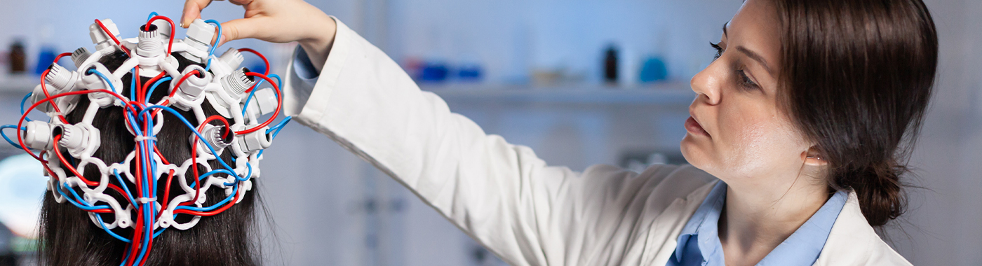
Neurons communicate using electrical signals. The summed activity of large populations of cortical neurons produces small voltage changes that can be detected on the scalp. During an EEG, small metal electrodes are fixed to the scalp with conductive gel, paste or a cap. The signals are amplified and displayed as continuous waveforms.
The reporting doctor looks at the background rhythm, its symmetry and reactivity, the response to eye opening and closing, sleep patterns if sleep is achieved, and whether any epileptiform discharges (spikes, sharp waves, spike-wave complexes) are present. Frequency bands, delta, theta, alpha, beta and gamma, are descriptive labels used in that analysis. They are not scores of brain health, and no single band tells you whether a person is intelligent, stressed or well.
Why an EEG is done
- Suspected seizures: to look for epileptiform abnormalities and help classify the seizure type and epilepsy syndrome.
- Classification and treatment choice: distinguishing generalised from focal epilepsy influences drug selection.
- تغير الوعي أو الارتباك: to identify encephalopathy, and to detect non-convulsive seizures or non-convulsive status epilepticus in ICU patients.
- Recurrent unexplained events: to help separate epileptic seizures from syncope, functional (dissociative) seizures, parasomnias or movement disorders, usually best answered by video EEG capturing an event.
- Suspected encephalitis, including herpes simplex encephalitis, where certain patterns can be supportive.
- اضطرابات النوم, as one channel within full polysomnography rather than as a stand-alone EEG.
- التقييم قبل الجراحة in drug-resistant epilepsy, where prolonged video EEG helps localise the seizure onset zone.
- Brain death assessment, only as an ancillary test in specific protocols; the determination is clinical and legally defined.
من ينبغي أن يخضع لهذا الاختبار؟
- Anyone who has had a first unprovoked seizure, as advised by a neurologist.
- People with recurrent blackouts, staring spells, unexplained jerks or episodes of unresponsiveness.
- Children with suspected absence epilepsy, infantile spasms, or regression of milestones with suspected epileptic activity.
- Patients with known epilepsy whose seizure pattern changes or who are being considered for stopping medication.
- ICU patients with unexplained coma or fluctuating consciousness.
- People being assessed for epilepsy surgery.
An EEG is not a general health screen. It is not useful for headache alone, giddiness alone, memory complaints alone, or as a routine check-up, and a normal EEG in those situations provides very little reassurance about anything specific.
What an EEG cannot detect or exclude
- It cannot exclude epilepsy. A substantial proportion of people with epilepsy have a normal first routine EEG. Repeat recordings, sleep-deprived studies or prolonged video EEG increase the chance of capturing abnormalities, but even repeated normal EEGs do not exclude the diagnosis.
- It cannot confirm epilepsy on its own. Some non-specific or "borderline" patterns, and some benign variants, occur in people who never have seizures. Over-reading of such patterns is a well-recognised cause of people being wrongly labelled epileptic.
- It does not show brain structure. Tumours, strokes, haemorrhage, tuberculomas, neurocysticercosis and cortical malformations require MRI or CT. A normal EEG does not mean the brain scan will be normal, and vice versa.
- It mostly sees the surface. Activity arising deep in the brain, in mesial temporal structures, or in small areas may not reach the scalp electrodes.
- It does not diagnose dementia, ADHD, autism, learning difficulty or psychiatric illness. The EEG has no established role as a diagnostic test for these conditions, and claims that brain-wave testing can diagnose them are not supported.
- It does not assess the heart. Fainting caused by arrhythmia needs cardiac evaluation, not an EEG.
- It does not measure blood flow or oxygen delivery.
- A negative finding is not the same as exclusion. "No epileptiform discharges seen" describes the recorded period only. If symptoms continue despite a normal report, return to your doctor rather than assuming the problem is settled.
فحوصات ذات صلة ولكنها مختلفة
| اختبار | السؤال يجيب عليه | الفرق الرئيسي |
|---|---|---|
| Routine (awake) EEG | Are epileptiform discharges or background abnormalities present in a short recording? | Shortest and most available; lowest yield |
| Sleep-deprived / awake-sleep EEG | Do discharges appear in drowsiness and sleep? | Higher yield than routine awake EEG; needs partial sleep restriction, which itself can provoke seizures in some people |
| Prolonged video EEG (in-hospital) | What exactly happens electrically during the patient's actual event? | Correlates behaviour on video with EEG; the main tool for separating epileptic from functional seizures and for surgical localisation |
| EEG متنقل | Can events be captured over days at home? | No video in many set-ups, so behaviour cannot be correlated as reliably |
| تخطيط النوم (دراسة النوم) | Is there sleep apnoea or another primary sleep disorder? | Includes EEG plus breathing, oxygen, ECG, limb and eye channels |
| Intracranial / invasive EEG | Precisely where does the seizure begin? | Surgical placement of electrodes; carried out only in epilepsy surgery programmes and carries operative risk |
| MRI brain (epilepsy protocol) | Is there a structural cause? | Anatomy, not electrical function, complementary to EEG, not a substitute |
| ECG / Holter | Is the heart rhythm causing the blackouts? | Records the heart; often the more appropriate first test for classic faints |
| Evoked potentials (VEP, BAER, SSEP) | Is a specific sensory pathway conducting normally? | Measures responses to stimuli, not spontaneous brain activity |
Which test comes first depends on the presentation. For a witnessed convulsion, EEG and MRI are usually both arranged. For a blackout with sweating, nausea and rapid full recovery while standing, cardiac and postural assessment usually comes first. For snoring with daytime sleepiness, polysomnography is the relevant study, not a routine EEG.
كيفية تحضير
Preparation is operational and differs between hospitals and EEG laboratories. The written instruction you are given by your own unit takes precedence over any general advice.
- شعر: wash hair the night before or on the morning of the test and avoid oil, gel, serum, wax or hair spray, as these stop electrodes sticking. Many Indian units specifically ask for no coconut or other hair oil.
- المنشطات: many units ask you to avoid coffee, tea, cola, energy drinks and nicotine for several hours before the study. The exact window is set by the unit.
- النوم: some studies are deliberately sleep-deprived; others require a normal night's sleep. Follow only what your unit has asked, as the two instructions are opposite.
- أدوية: continue all prescribed medicines, including anti-seizure drugs, unless the neurologist who ordered the EEG has told you otherwise in writing. Never stop or reduce an anti-seizure medicine on your own, withdrawal can provoke seizures, including status epilepticus. Bring a current list of all medicines, including AYUSH, herbal and over-the-counter products.
- الطعام: fasting is not usually required, and going without food can alter the recording. Ask your unit if unsure.
- احضر: a description of your events, ideally with a mobile phone video recorded by a family member, plus previous EEG and MRI reports. Event videos are often more informative than the EEG itself.
- مرافقة: bring a relative if you have frequent seizures, are sleep-deprived, or if a child is being tested. Do not drive yourself to a sleep-deprived EEG.
أطفال
Paediatric EEG may be scheduled around nap time. Feeding, sleep timing and whether any sedation is used are decided by the paediatric neurology team. Bring a favourite toy or blanket, and a spare set of clothes for infants.
فترة الحمل
EEG involves no radiation and is performed in pregnancy when clinically indicated. Sleep deprivation and hyperventilation protocols may be modified, and anti-seizure medicines must not be changed without the prescriber's advice.
مرضى السكري الذين يتلقون علاجًا لخفض مستوى الجلوكوز
If any fasting or meal timing change is requested, tell the unit that you take insulin or other glucose-lowering medicines before the appointment, so timings and doses can be planned with your prescriber. Low blood glucose can itself alter the EEG and cause symptoms.
Older adults, heart failure and advanced CKD
Lying flat for a prolonged recording can be uncomfortable if you are breathless, and prolonged studies may need positioning adjustments. Fluid-restricted patients should not increase fluid intake for any test instruction without checking with the treating doctor. Tell the unit about mobility limitations, oxygen use or dialysis schedules.
Scalp conditions and hair styling
Open wounds, severe eczema, recent scalp surgery, thick braids, extensions, weaves or dreadlocks can limit electrode placement. Inform the unit in advance so an alternative approach can be planned.
What happens during an EEG
- القياس ووضع العلامات: the technologist measures your head and marks electrode positions using the 10-20 system.
- Electrode application: the scalp is lightly cleaned and electrodes are fixed with gel or paste, or a cap is applied. This can feel cool and slightly scratchy but is not cutting or piercing.
- تسجيل: you lie or sit still with eyes closed for most of the study, opening and closing the eyes on request.
- Activation procedures: hyperventilation (deep breathing) and photic stimulation (a flashing light) are commonly used to bring out abnormalities. These are done selectively and can occasionally provoke a seizure, that is a recognised part of the test, and staff are prepared for it. Tell staff if you have known photosensitivity, severe respiratory disease, recent stroke or sickle cell disease before hyperventilation or flashing lights are used.
- Sleep recording: you may be encouraged to fall asleep, since discharges often appear in drowsiness and sleep.
- عقب ذلك مباشرة: electrodes are removed and residual gel or paste is cleaned off. Hair may feel sticky and usually needs washing at home. Most people resume normal activity immediately.
كم من الوقت تستغرق العملية
- تخطيط كهربية الدماغ الروتيني: typically around 20-40 minutes of recording, with roughly 15-30 minutes extra for electrode application and removal.
- Awake-sleep or sleep-deprived EEG: often 1-2 hours, sometimes longer if sleep is slow to come.
- تخطيط كهربية الدماغ المتحرك: usually 24 hours to several days, with equipment worn at home.
- In-hospital video EEG: commonly several days of admission, continuing until enough events or enough sleep-wake cycles are recorded.
Reporting time varies between units, since an EEG must be read by a trained physician and interpreted against your clinical details. Ask the unit when and how you will receive the report.
Understanding your EEG report
An EEG report usually describes the recording conditions, the background activity, any asymmetry or slowing, whether sleep was achieved, the response to activation procedures, and whether epileptiform discharges were seen. It ends with an interpretation.
- "Normal EEG": the background was age-appropriate and no abnormal discharges were recorded during that period. The EEG was normal, you were not necessarily proven free of epilepsy.
- Epileptiform discharges (spikes, sharp waves, spike-wave): support an epilepsy diagnosis and help classify it, but must fit the clinical picture.
- Focal slowing: suggests a localised disturbance of function. It is non-specific and can follow a seizure, a structural lesion, or drug effect. It usually prompts imaging rather than a diagnosis by itself.
- Generalised slowing: commonly reflects encephalopathy from metabolic, infective, toxic or drug causes, or drowsiness during the recording. It is not a measure of intelligence or of "brain damage" in any general sense.
- "Borderline", "mild non-specific abnormality", "benign variant": these phrases are frequently over-interpreted by patients. Many such patterns have no clinical significance. Ask the neurologist what, if anything, it changes.
- Electroclinical seizure: a seizure was recorded, with or without visible symptoms.
An EEG never diagnoses on its own. Results must be interpreted in clinical context by the doctor who requested the test, and results vary depending on individual clinical circumstances.
When EEG results can be misleading
Falsely suggesting abnormality (false positives)
- Muscle activity, chewing, swallowing, eye or eyelid movement, sweating and electrode "pop" artefact can mimic abnormalities.
- Mains interference, mobile phones and nearby equipment create electrical artefact.
- Benign normal variants are sometimes misread as epileptiform.
- Normal sharp transients of drowsiness and sleep can be over-read.
- Reporting without clinical details increases the risk of over-diagnosis, which can lead to unnecessary years of anti-seizure medication and avoidable driving or employment restrictions.
Falsely suggesting normality (false negatives)
- Short recordings that do not capture sleep frequently miss discharges.
- Infrequent epilepsy may have long discharge-free intervals.
- Deep or small generators may not project to the scalp.
- Anti-seizure medicines, sedatives and benzodiazepines can suppress discharges.
- Poor electrode contact from hair oil, styling products or a sweaty scalp degrades the signal.
- An excessively drowsy or restless patient, or a child who will not settle, yields a limited study.
Direction of error with drugs and physiology
- Sedatives, alcohol and anaesthetics: slow the background and can mask discharges.
- Caffeine and alertness: reduce drowsiness, lowering the chance of capturing sleep-related discharges.
- Hypoglycaemia, hyponatraemia, uraemia, liver failure, hypoxia, fever: cause slowing that may be misattributed to a primary brain disorder.
- Recent seizure: can produce transient post-ictal slowing that resolves.
المخاطر والسلامة
Routine scalp EEG
A routine scalp EEG is non-invasive, uses no radiation and does not pass current into the head. Common issues are minor: pressure or scratchiness during electrode placement, sticky hair afterwards, and occasional mild skin irritation or contact reaction from gel, paste or adhesive. Rarely, minor skin abrasion occurs where the scalp is prepared.
Activation procedures
Hyperventilation can cause tingling, light-headedness and hand cramping, which settle when normal breathing resumes. Photic stimulation may trigger a seizure in photosensitive individuals. In people with epilepsy, either procedure can provoke a seizure; this is sometimes the intended diagnostic outcome and is managed by trained staff.
Sleep-deprived EEG
Sleep restriction can itself provoke seizures and causes daytime sleepiness. Arrange transport rather than driving.
Prolonged video EEG monitoring
Because medication may be reduced under supervision to capture events, there is a higher risk of seizures, including clusters, injury during a seizure, or status epilepticus. This is why such monitoring is carried out as an inpatient with continuous observation. Skin breakdown under long-term electrodes can also occur.
Sedation in children
Where sedation is used, it carries its own risks relating to breathing and airway, and is given only with appropriate monitoring by staff trained for it.
تخطيط كهربية الدماغ داخل الجمجمة
Invasive electrode placement is a neurosurgical procedure with risks including bleeding, infection and neurological deficit. It is undertaken only in specialised epilepsy surgery programmes after detailed counselling. Risk profiles differ substantially across these EEG types and cannot be described with one blanket safety statement.
الأعلام الحمراء
Emergency now: call an ambulance or go to the nearest emergency department
- A seizure lasting more than five minutes, or repeated seizures without recovery in between.
- Seizure followed by failure to regain consciousness.
- Difficulty breathing, blue lips, or persistent vomiting after a seizure.
- First-ever seizure, especially with head injury, fever with neck stiffness, or pregnancy.
- New weakness of one side, loss of speech, or sudden severe headache.
- Seizure in a person with diabetes on insulin or other glucose-lowering treatment, where hypoglycaemia is possible.
- Seizure occurring in water, causing significant injury, or with suspected poisoning or overdose.
Same day: contact your neurologist or attend urgent care today
- A clear increase in seizure frequency or a new seizure type.
- Seizures after a missed or changed dose, or after a new medicine was started.
- New confusion, drowsiness or behavioural change lasting hours.
- Rash, fever or mouth ulcers after starting an anti-seizure medicine.
- Fever, headache and altered behaviour with suspected encephalitis.
موعد روتيني
- Collecting and discussing your EEG report.
- Persisting symptoms despite a normal EEG.
- Unexplained staring or blank spells, brief jerks on waking, or memory gaps.
- Mild scalp irritation that is settling.
- Questions about driving, work safety, contraception, pregnancy planning or stopping medication, discuss with the prescriber; do not stop treatment yourself.
حالات خاصة
فترة الحمل
EEG is safe with respect to radiation because none is used, and is performed during pregnancy when indicated. Anti-seizure medication decisions in pregnancy are specialised and should involve both the neurologist and the obstetrician. Do not alter doses independently.
Children and neonates
Paediatric EEG interpretation depends heavily on age, since normal background changes with maturation. Neonatal EEG is a distinct specialist study. Certain childhood syndromes have characteristic EEG patterns, which is why EEG is often more informative in children than in adults.
كبار السن
Slowing may reflect medication, drowsiness or systemic illness rather than a primary brain disease. EEG is not a test for dementia, though it may help when seizures or encephalopathy are suspected in someone with cognitive decline.
People with diabetes on glucose-lowering therapy
Blood glucose should not be allowed to fall for the sake of a test. If sleep deprivation or meal timing changes are requested, plan them with the prescriber.
مرض الكلى المزمن وفشل القلب
Uraemia and metabolic disturbance produce non-specific slowing that can be misread. Prolonged supine recording may be difficult with breathlessness. Never increase fluid intake for a test instruction if you are on a fluid restriction.
ICU and post-cardiac-arrest patients
Continuous EEG is used to detect non-convulsive seizures and to help with prognostication after cardiac arrest. Sedatives, temperature control and organ failure all influence the recording, so findings are interpreted cautiously and serially rather than from a single tracing.
الغرسات والأجهزة
Cardiac pacemakers, deep brain stimulators and vagus nerve stimulators do not prevent an EEG, though they may introduce artefact. Tell the technologist about any implanted device. If MRI is also planned, MRI safety assessment of the device is required separately.
EEG in the Indian context
- Common local causes of seizures matter for workup. In India, neurocysticercosis and tuberculoma are important structural causes of focal seizures. An EEG cannot show either; MRI or CT of the brain is needed. A normal EEG with a new focal seizure should not stop imaging.
- Terminology confusion is frequent. Patients often arrive asking for a "brain test" or confuse EEG with ECG ("heart test"), EMG/NCV (nerve and muscle test) and MRI. Confirm on the prescription which test has been ordered.
- Hair oil is a practical problem. Coconut and other hair oils, and heavy styling products, commonly degrade electrode contact and can force a repeat study. Attend with clean, dry, product-free hair unless told otherwise.
- Stigma and misinformation. Epilepsy still attracts social stigma in parts of India, and some people are offered unproven "brain wave" or "neuro-scan" treatments. Under the Drugs and Magic Remedies (Objectionable Advertisements) Act 1954, advertisements claiming cures for epilepsy are not permitted. Treatment decisions belong with a qualified neurologist or physician.
- Medication access and continuity. Interrupted supply of anti-seizure medicines is a common reason for breakthrough seizures. Plan refills ahead of travel and festivals, and speak to the prescriber rather than stopping treatment.
- Sleep-deprived EEG and travel. Long commutes on the day of a sleep-deprived study increase the risk of a seizure or accident in transit. Arrange for someone to accompany you.
- يختلف التوفر. Routine EEG is widely available; prolonged video EEG, ambulatory EEG and epilepsy surgery evaluation are concentrated in larger centres, often requiring referral.
التكاليف والتأمين في الهند
Costs differ widely between government facilities, trust hospitals, standalone diagnostic centres and corporate hospitals, and between cities. Rather than quoting a figure, it is more useful to know what drives the price. Always ask for a written estimate before booking.
- Type of EEG: a routine 20-40 minute study costs far less than a sleep-deprived, ambulatory or multi-day inpatient video EEG.
- Duration and channels: longer recordings and higher-density electrode arrays cost more.
- Video and inpatient component: prolonged video EEG includes bed charges, nursing and continuous observation.
- Sedation or anaesthesia support in young children adds cost.
- التقرير: specialist neurologist or epileptologist reporting, and urgent or after-hours reporting, may be charged separately.
- City and sector: metro private centres are generally costlier than smaller-town or public facilities.
- التجميع: EEG is often part of a package with MRI brain and blood tests during a seizure workup.
التأمين: outpatient diagnostics are often not covered under basic indemnity health insurance unless an OPD benefit applies. When EEG is performed during an admission, for example inpatient video EEG or evaluation of status epilepticus, it is usually considered part of the hospitalisation claim, subject to policy terms, waiting periods and pre-existing disease clauses. Obtain pre-authorisation where required and keep the prescription, reports and bills. Government schemes and state health schemes have their own listed rates and referral pathways.
الخرافات والحقائق
| أسطورة | حقيقة |
|---|---|
| A normal EEG means I do not have epilepsy. | Routine EEG is often normal in epilepsy. The diagnosis rests on the clinical history, supported by EEG and imaging. |
| An abnormal EEG means I definitely have epilepsy. | Some patterns are benign variants or artefact. The report must be read alongside your symptoms. |
| EEG passes electricity into the brain or gives shocks. | The EEG only records. Nothing is delivered into the head. This is different from ECT, which is a separate treatment. |
| EEG shows tumours and clots. | EEG shows function. Structural lesions need MRI or CT. |
| EEG measures intelligence, stress or personality. | It does none of these. There is no validated EEG score for intelligence or character. |
| EEG can diagnose ADHD, autism or dementia. | These are clinical diagnoses. EEG has no established stand-alone diagnostic role in them. |
| I should stop my seizure medicine before an EEG so the test shows the truth. | Never stop anti-seizure medicine yourself. Any reduction is done only when a specialist advises it, usually under supervision. |
| EEG involves radiation. | لا يتم استخدام الإشعاع. |
| My head will be shaved. | Shaving is not routine. Occasionally a very small area is cleared for long-term monitoring. |
الأسئلة المتكررة
هل تخطيط الدماغ الكهربائي مؤلم؟
An EEG is not painful. Electrodes are placed on the surface of the scalp with gel or paste, and nothing punctures the skin. You may feel mild scratchiness during scalp preparation, coolness from the gel, and pressure from a cap. Hair feels sticky afterwards and usually needs washing at home.
هل يمكن لجهاز تخطيط الدماغ الكهربائي (EEG) الكشف عن مشكلة في القلب؟
No. An EEG records brain activity, not heart rhythm. Many EEG set-ups include one ECG channel to identify heart-rate artefact, but that is not a cardiac assessment. If fainting may be cardiac in origin, an ECG, Holter monitoring or echocardiogram is the appropriate investigation.
Do I need to fast before an EEG?
Fasting is not usually required, and skipping meals can alter the recording and provoke symptoms, particularly in people taking insulin or other glucose-lowering medicines. Some units request that caffeine and nicotine be avoided for a few hours. Follow the written instruction from the unit performing your test.
Will one EEG be enough?
Sometimes, but not always. If the first routine EEG is normal and seizures are still suspected, the neurologist may request a sleep-deprived recording, a repeat study, or prolonged video EEG. Each additional recording increases the chance of capturing abnormal activity, though no number of normal studies fully excludes epilepsy.
Can an EEG trigger a seizure?
Occasionally, yes. Hyperventilation, flashing lights and deliberate sleep deprivation can provoke seizures, and in prolonged video EEG this is sometimes the aim. Trained staff monitor you throughout and manage any seizure that occurs. Tell the team beforehand if you know that flashing lights affect you.
Should I wash my hair before the EEG?
Yes, unless told otherwise. Clean, dry hair without oil, gel, serum, wax or spray allows electrodes to make proper contact. Oiled hair is a common reason for poor-quality recordings and repeat appointments. Braids, extensions and weaves may need to be loosened, so inform the unit in advance.
How is an EEG different from an MRI brain?
The two tests answer different questions and are often ordered together. EEG records electrical function over time and can show epileptiform activity. MRI shows brain structure and can reveal scars, tumours, infarcts, tuberculomas or malformations. Neither substitutes for the other, and a normal result in one does not predict the other.
Can children have an EEG safely?
Yes, EEG is widely used in children and involves no radiation. Recordings are often timed around sleep, and interpretation is age-specific because normal patterns change as the brain matures. Sedation is used only when necessary, with appropriate monitoring. Bring comfort items and a description or video of the events.
My report says "mild non-specific abnormality". Should I worry?
Not necessarily. Such wording often reflects drowsiness, artefact or a benign variant, and frequently changes nothing about management. It is not a diagnosis. Take the report to the doctor who ordered the test and ask sp









 أفضل مستشفى بالقرب مني في تشيناي


