


Respuesta directa
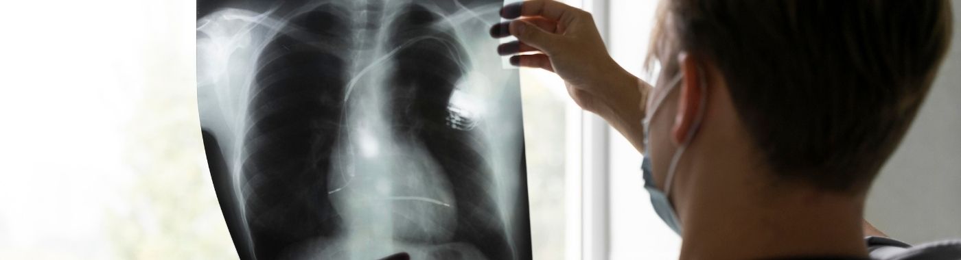
A chest X-ray is a quick imaging test that uses a small dose of ionising radiation to produce a two-dimensional picture of the lungs, heart outline, ribs, diaphragm and major airways. The chest X-ray helps assess breathlessness, cough, fever, chest injury and heart failure, but a normal chest X-ray does not exclude serious chest disease.
Puntos clave
- A chest X-ray is fast, painless and widely available, and is often the first imaging step for cough, fever, breathlessness or chest injury.
- A chest X-ray shows shadows and outlines, not tissue detail, it cannot visualise coronary arteries, cannot grade heart function, and can miss small lung nodules, early tuberculosis and many rib fractures.
- A normal chest X-ray means nothing abnormal was seen on that film, not that disease has been excluded.
- Radiation dose from a single chest X-ray is low, but the test should only be done when there is a clinical reason; routine screening X-rays without an indication are not advised.
- Findings such as "prominent bronchovascular markings" or "cardiomegaly on a portable film" are often non-specific and must be interpreted by the doctor who requested the test.
De un vistazo
| Elemento | Detail |
|---|---|
| Lo que muestra | Lungs, pleural spaces, heart and mediastinal outline, diaphragm, ribs, clavicles, upper spine, soft tissues of the chest wall |
| Tecnología | Ionising radiation (X-rays), digital or film radiography |
| Vistas estándar | PA (back-to-front, standing) and often a lateral view; AP (portable) when the patient cannot stand |
| Preparación | Usually none; no fasting required |
| Time in the X-ray room | Typically 2-5 minutes for image capture; 10-15 minutes including changing and positioning |
| Malestar | Painless; brief breath-hold and arm positioning |
| Contrast injection | No utilizado |
| Limitación de la clave | Two-dimensional summation image; overlapping structures hide lesions; low sensitivity for small or early disease |
También conocido como
- CXR: the standard clinical abbreviation
- Radiografía de tórax: the formal radiology term
- X-ray chest PA view: the most commonly ordered request in Indian laboratories
- Chest film, placa pectoral: still used colloquially in many Indian hospitals from the film era
- Seene ka X-ray (Hindi), maarbu X-ray (Tamil), chaati ka X-ray: common patient terms
What a chest X-ray is
A chest X-ray passes a controlled beam of X-rays through the chest onto a digital detector. Dense structures such as bone absorb more of the beam and appear white; air-filled lung appears dark; heart, vessels and fluid appear in shades of grey. The resulting image is a flat summation of everything the beam passed through, which is why a chest X-ray gives excellent overall orientation but limited fine detail.
Two standard views are common. The PA view is taken with the patient standing, chest against the detector, and gives the truest heart size. The vista lateral adds depth and helps find lesions hidden behind the heart or diaphragm. A portable AP film, taken at the bedside in sick or immobile patients, magnifies the heart shadow and is less reliable for judging heart size or small pleural fluid.
Why a chest X-ray is done
- Síntomas respiratorios: persistent cough, fever, breathlessness, coughing up blood, or suspected pneumonia, tuberculosis or lung collapse.
- Evaluación cardíaca: to look for an enlarged cardiac silhouette, pulmonary congestion or pleural effusion in suspected heart failure.
- Chest injury: to look for pneumothorax, haemothorax or displaced rib fractures after trauma.
- Monitoreo: tracking resolution of pneumonia, response to anti-tubercular therapy, or progress after chest surgery.
- Device and tube position: confirming placement of endotracheal tubes, central lines, chest drains and pacemaker leads.
- Occupational and pre-employment screening: for suspected occupational lung disease such as silicosis, or where an employer or visa authority requires it.
- Cuerpo extraño: when inhalation or ingestion of a radio-opaque object is suspected.
¿Quién debería hacerse esta prueba?
- Adults with cough lasting more than two weeks, especially with fever, weight loss or night sweats, a common tuberculosis evaluation pathway in India.
- Anyone with new or worsening breathlessness where the cause is unclear.
- People with chest trauma, or sudden sharp chest pain with breathlessness where pneumothorax is suspected.
- Patients with known heart failure or COPD having a flare-up.
- Patients being assessed before major surgery, when the anaesthetist or surgeon requests it.
- People starting anti-tubercular therapy or certain immunosuppressive medicines, when the treating doctor advises baseline imaging.
A chest X-ray is not recommended as a routine annual test in a person with no symptoms and no risk factors. Repeated imaging without an indication adds radiation exposure without adding benefit. Low-dose CT, not chest X-ray, is the modality studied for lung cancer screening in high-risk smokers; chest X-ray has not been shown to reduce lung cancer deaths when used as a screening test.
What a chest X-ray cannot detect or exclude
This is the single most misunderstood aspect of the test. A chest X-ray is a screening-level overview, not a definitive answer.
- Enfermedad de la arteria coronaria: the chest X-ray does not visualise coronary arteries. A normal chest X-ray says nothing about whether the arteries are blocked and does not exclude a heart attack or angina.
- Embolia pulmonar: a chest X-ray cannot confirm or exclude a clot in the lung arteries. CT pulmonary angiography is the test for that question.
- Small lung nodules and early lung cancer: lesions under roughly a centimetre, and lesions hidden behind the heart, diaphragm, hila or ribs, are frequently invisible. A normal chest X-ray does not exclude lung cancer.
- Early or minimal tuberculosis: a chest X-ray may look normal in early pulmonary TB and cannot distinguish active from healed disease with confidence. Diagnosis needs sputum testing such as smear microscopy and nucleic acid amplification (for example, CBNAAT/NAAT), plus clinical assessment.
- Función cardíaca: a chest X-ray cannot measure ejection fraction, assess valves or detect rheumatic valve disease reliably. An echocardiogram is required.
- Many rib fractures: undisplaced rib fractures and costal cartilage injuries are often not seen. A normal film does not exclude a fractured rib.
- Interstitial lung disease in early stages: high-resolution CT is far more sensitive.
- Asma: a chest X-ray in asthma is usually normal. Asthma is diagnosed with spirometry and clinical history, not imaging.
- Small pleural effusions and small pneumothoraces: particularly on supine or portable films.
- Oesophageal and mediastinal detail: soft tissue characterisation needs CT or MRI.
If a chest X-ray is reported as normal but symptoms continue or worsen, return to your doctor. A normal report describes what was visible on that film; it does not rule out disease.
Exámenes relacionados pero diferentes
| Prueba | Pregunta que responde | How it differs from a chest X-ray |
|---|---|---|
| Chest CT (plain) | Is there a nodule, mass, interstitial disease or subtle infection? | Cross-sectional slices, far greater sensitivity, higher radiation dose, higher cost |
| angiografía pulmonar por tomografía computarizada | Is there a pulmonary embolism? | Uses iodinated contrast; requires kidney function and allergy assessment |
| High-resolution CT (HRCT) chest | What is the pattern of interstitial lung disease? | Thin-section protocol targeted at lung architecture |
| Low-dose CT | Screening for lung cancer in defined high-risk groups | Lower dose than standard CT; chest X-ray is not an equivalent screening test |
| Echocardiogram (echo test) | How well does the heart pump, and are the valves normal? | Ultrasound, no radiation; assesses function, which X-ray cannot |
| Lung/pleural ultrasound | Is there pleural fluid, and where should it be drained? | No radiation, bedside, better than X-ray for small effusions; cannot see through aerated lung |
| resonancia magnética de tórax | Detailed soft-tissue and chest wall assessment | No ionising radiation; limited for lung parenchyma; requires MRI safety assessment for implants |
| Fluoroscopy / screening of diaphragm | Is the diaphragm moving normally? | Real-time moving images rather than a still frame |
| Sputum testing for TB | Is Mycobacterium tuberculosis ¿regalo? | Microbiological, not imaging; a chest X-ray can suggest TB but cannot confirm it |
Which test comes first depends entirely on the clinical problem. For sudden pleuritic chest pain with breathlessness, a chest X-ray may come first to look for pneumothorax, but CT pulmonary angiography answers the embolism question. For exertional chest pain, cardiac assessment, not a chest X-ray, is the relevant pathway. For chronic cough with weight loss in India, sputum testing runs alongside imaging rather than after it.
How to prepare for a chest X-ray
Preparation is minimal, but instructions vary between units. Follow the specific instructions given by the imaging department where your scan is booked, those take precedence over any general guidance.
- No fasting: eat and drink normally. Take your regular prescribed medicines as usual unless your prescriber tells you otherwise.
- Ropa: wear something without metal fasteners above the waist. You may be asked to change into a gown.
- Eliminar metal: necklaces, hair clips, bra with underwiring, coins in pockets and some medicated patches can create shadows that mimic or hide disease.
- Trae películas antiguas: comparison with a previous chest X-ray is one of the most valuable things a radiologist can have. Carry earlier films, CDs or reports.
- Tell the staff: if you are or may be pregnant, if you have had recent chest imaging, or if you cannot stand or hold your breath.
If you may be pregnant
Tell the radiographer before the examination. A chest X-ray delivers a very low dose and the uterus is outside the primary beam, so a clinically necessary chest X-ray is often still performed during pregnancy after discussion. The decision rests with your treating doctor and the radiologist, weighing the risk of missing a diagnosis against the small radiation exposure.
Niños
Paediatric chest X-rays use reduced exposure settings. A parent may be asked to help with positioning and will be given protective wear. Immobilisation aids are sometimes used in infants to avoid repeat films from movement.
People who cannot stand
A portable AP film is taken sitting up in bed or lying down. Image quality is lower, the heart appears larger than it truly is, and small effusions or pneumothoraces are easier to miss. The report will usually say the film is portable or AP, this context matters when comparing with earlier films.
Older adults and people with breathing difficulty
Holding a breath in full inspiration may be hard. Tell the radiographer; the exposure can be timed to your breathing. A film taken in poor inspiration commonly produces crowded lung markings and an apparently enlarged heart, and can be misread as abnormal.
What happens during a chest X-ray
- posicionamiento: for a PA view you stand facing the detector, chin raised, hands on hips or arms wrapped forward to move the shoulder blades out of the lung fields.
- Breath-hold: you take a deep breath in and hold it for two to three seconds while the image is taken.
- Second view: a lateral view may follow, with your arms raised and your side against the detector.
- Checking the image: the radiographer reviews the image immediately and may repeat it if movement or positioning has degraded it.
- Después: there is no recovery period and no after-effects. Normal activity resumes at once.
¿Cuánto tiempo tarda el proceso?
The X-ray exposure itself lasts a fraction of a second. Image capture for a PA view takes about 2-5 minutes including positioning; adding a lateral view or waiting for a gown change usually brings the total to 10-15 minutes. Waiting time before the examination and reporting time afterwards depend on the unit's workload, ask at the reception desk for their current timings, as urgent and inpatient films are typically reviewed sooner than routine outpatient films.
Understanding your chest X-ray report
A chest X-ray report is descriptive. The radiologist comments on lung fields, heart size and shape, mediastinum, hila, pleura, diaphragm, bones and soft tissues, then gives an impression. There is no numeric "normal range" for a chest X-ray, and any source presenting one is misleading, interpretation is visual and comparative.
Common phrases and what they actually mean
- "Both lung fields are clear": no visible consolidation, mass or fluid on this film. It is not a statement that the lungs are healthy.
- "Cardiothoracic ratio within normal limits": the heart shadow is not enlarged on this view. Valid only on a properly taken PA film, not on a portable AP film.
- "Prominent bronchovascular markings": a frequently reported, non-specific description. It can reflect a shallow breath, normal variation, bronchitis or congestion. On its own it is not a diagnosis.
- "Costophrenic angle blunting": suggests pleural fluid or old pleural thickening. Ultrasound of the chest usually clarifies which.
- "Fibrotic/calcified opacity in the upper zone": often signifies old, healed tuberculosis, extremely common in India. It does not mean active TB, and treatment is not started on this finding alone.
- "Hyperinflated lungs with flattened diaphragms": can suggest airflow obstruction such as COPD, but needs spirometry for confirmation.
- "Suggest clinical correlation" or "suggest CT for further evaluation": the radiologist has seen something that a flat image cannot characterise. This is routine, not alarming in itself.
Results vary depending on individual clinical circumstances. No chest X-ray finding should be self-interpreted. The doctor who requested the test will read the report alongside your symptoms, examination and other tests before deciding anything.
When chest X-ray results can be misleading
Findings that can appear falsely abnormal
- Poor inspiration: a shallow breath crowds the lung markings and widens the heart shadow, imitating cardiomegaly and pulmonary congestion.
- Portable AP films: magnify the heart and mediastinum; a normal heart may be reported as enlarged.
- Rotación: a rotated patient can create apparent mediastinal shift or one-sided lung darkening.
- Overlying objects: buttons, hair plaits, nipple shadows, ECG leads and skin lesions can mimic lung nodules.
- Breast implants, chest wall fat or thick soft tissues: can create hazy lower zones resembling consolidation.
- Old healed scarring: can be misread as new disease if no previous film is available for comparison.
Findings that can appear falsely normal or be underestimated
- Zonas ocultas: lesions behind the heart, in the lung apices, at the hila, or below the diaphragm are easily missed on a single PA view.
- Early pneumonia: consolidation may not be visible in the first hours of illness or in dehydrated patients, appearing only on a repeat film.
- Neutropenia or immunosuppression: infection may produce little visible change despite serious illness.
- Supine films: pleural fluid layers posteriorly and pneumothorax collects anteriorly, so both can be invisible when the patient is lying flat.
- Obesity and high body thickness: reduce contrast resolution and can obscure subtle findings.
- Digital post-processing: heavy image processing can occasionally soften genuine abnormalities.
Riesgos y seguridad
Standard chest X-ray
A chest X-ray uses one of the lowest radiation doses of any medical imaging examination, broadly comparable to a few days of ordinary background radiation, though exact dose varies with equipment and patient size. There is no injection, no sedation and no recovery period. The main consideration is avoiding unnecessary repeat exposure, which is why previous films should always be brought along. Radiation risk is cumulative over a lifetime, so each examination should have a clinical reason.
Portable and repeated films in hospital
Inpatients, particularly in intensive care, may have daily films. Each has a low individual dose, but the department will limit them to those that change management. Image quality is lower than departmental films.
Embarazo
The foetus is not in the direct beam during a chest X-ray and the scattered dose is very small. Even so, the examination is performed in pregnancy only when the information is needed. Discuss alternatives such as chest ultrasound with your doctor where appropriate.
Niños
Children are more radiosensitive than adults and have longer life expectancy for any effect to appear, so paediatric protocols use reduced settings and tight collimation. A chest X-ray in a child is still justified where clinically indicated.
Red flags: when to act on chest symptoms
Emergency now: call emergency services or go to the nearest emergency department
- Severe breathlessness, inability to speak in full sentences, or blue lips or fingertips
- Crushing, heavy or pressure-like chest pain, particularly with sweating, nausea or pain spreading to the arm or jaw
- Sudden sharp one-sided chest pain with severe breathlessness after injury or spontaneously
- Coughing up a large amount of blood
- Collapse, confusion, or a chest injury after a significant fall or road accident
El mismo día: contacte hoy mismo con su médico.
- Fever with cough and breathlessness
- Small amounts of blood in the sputum
- New chest pain that worsens on breathing
- Rapidly increasing swelling of both legs with breathlessness
- A report advising urgent review, or a phone call from the imaging department
Cita de rutina
- Cough persisting beyond two to three weeks without alarming features
- An incidental finding such as an old calcified scar or mild scoliosis on a report
- A request in the report to correlate clinically or to obtain a comparison film
- Gradually reducing exercise tolerance over months
Situaciones especiales
Embarazo
A clinically indicated chest X-ray, for example to investigate suspected pneumonia or tuberculosis, may be performed at any stage of pregnancy after a discussion of risks and benefits. Tell the department before the examination so appropriate technique can be used.
Niños
Chest X-rays in children are commonly requested for suspected pneumonia, inhaled foreign body or persistent wheeze. Routine chest X-rays are not needed for every childhood fever or for uncomplicated asthma. The thymus can cause a normal widened upper mediastinal shadow in infants, which is sometimes mistaken for a mass.
Adultos mayores
Spinal curvature, previous fractures and reduced mobility affect positioning and image quality. Comparison with earlier films becomes especially important, since many older adults have long-standing changes that are stable and harmless.
People living with heart failure
Chest X-ray can show pulmonary congestion and pleural effusion and is useful for tracking fluid status, but it cannot measure pumping function. Echocardiography remains the test for that. Findings may lag behind clinical improvement.
People with COPD or long-standing lung disease
Chronic changes can mask new infection. A previous baseline film is the most helpful comparison available.
People on anti-tubercular therapy
Radiological improvement is slower than clinical improvement, and residual scarring often persists permanently. Do not interpret a persisting shadow as treatment failure, that judgement belongs to your treating physician, who will also use sputum results. Never stop or alter anti-tubercular medication on the basis of an X-ray report; speak to the prescriber.
People with pacemakers, stents or implants
Implanted devices do not prevent a chest X-ray; the test is often used to check lead position. Concerns about metal implants apply to MRI, which requires an MRI safety assessment, not to X-ray.
Chest X-ray in the Indian context
- Tuberculosis: India carries a substantial share of the global TB burden, and chest X-ray is a core tool in the National TB Elimination Programme, increasingly used for triage alongside molecular testing. Chest X-ray alone cannot confirm TB or distinguish active from healed disease; sputum-based microbiological confirmation is required before treatment. For current national figures, refer to the WHO Global Tuberculosis Report and the Government of India TB annual report for the relevant year.
- Old healed TB scarring: upper-zone fibrosis and calcified nodes are very common findings on routine Indian chest X-rays, frequently in people who never knew they had an infection. These findings often need no treatment, only correlation.
- Exposición ocupacional: silicosis and coal-worker's lung disease remain relevant in mining, stone-crushing and construction. Chest X-ray with an occupational history is central to assessment.
- Air pollution and biomass smoke: chronic respiratory symptoms are common in both urban and rural households, and chest X-ray helps distinguish structural disease from symptoms needing spirometry.
- Cardiopatía reumática: still prevalent in parts of India. A chest X-ray may hint at chamber enlargement or pulmonary congestion, but valve disease is diagnosed with an echo test, not a chest film.
- Reglamento: diagnostic X-ray installations in India operate under Atomic Energy Regulatory Board (AERB) requirements for equipment registration, room shielding and radiation safety. Radiographers and radiologists work to these standards. Note that PC-PNDT restrictions apply to ultrasound and prenatal sex determination, not to chest radiography, but sex determination in any form remains illegal.
- Protección de Datos: images and reports are personal health data handled under the Digital Personal Data Protection Act 2023.
- Visa, employment and insurance screening: many Indian employers and overseas authorities request a chest X-ray. Such a film is a screening document and does not replace clinical evaluation of symptoms.
Costo y seguro en India
A chest X-ray is among the least expensive imaging tests available in India. Indicative outpatient charges commonly fall in the low hundreds of rupees for a single PA view in public and smaller private facilities, with higher charges in large corporate hospitals and for additional views. Costs are indicative only and change frequently, confirm the current price with the facility before booking.
Factores que influyen en el costo:
- Number of views (PA only versus PA plus lateral, or additional oblique views)
- Digital radiography versus computed radiography versus conventional film
- Portable bedside film versus departmental film
- Whether a formal radiologist's report is included or charged separately
- Outpatient versus inpatient or emergency setting
- City tier and whether the facility is government, trust-run or corporate
- Whether hard-copy film, CD or digital transfer is requested
Seguro: outpatient diagnostic tests are often not covered under standard indemnity health insurance unless the policy includes an OPD benefit. A chest X-ray done as part of an admitted hospital stay is usually covered within the hospitalisation claim. Pre-employment and visa screening X-rays are generally not reimbursable. Government schemes such as Ayushman Bharat PM-JAY cover diagnostics linked to eligible admissions at empanelled hospitals. Check your specific policy wording.
Mitos y realidades
| Myth | Hecho |
|---|---|
| A normal chest X-ray means my lungs and heart are healthy | A normal film means nothing abnormal was visible. It does not exclude coronary disease, pulmonary embolism, small cancers or early TB. |
| A chest X-ray can diagnose tuberculosis | A chest X-ray can raise suspicion of TB. Confirmation needs sputum microbiological testing. Old scars are often mistaken for active disease. |
| A chest X-ray shows blocked heart arteries | Coronary arteries are not visible on a chest X-ray. Cardiac assessment requires other tests. |
| One X-ray causes serious radiation harm | The dose from a single chest X-ray is very low. The sensible approach is avoiding unnecessary repeats, not refusing indicated imaging. |
| Everyone should have a yearly chest X-ray | Routine chest X-rays in people without symptoms or risk factors are not recommended and have not been shown to save lives as a screening test. |
| An enlarged heart on the report means heart failure | Heart size is easily exaggerated by a portable film or a shallow breath. Confirmation requires an echocardiogram. |
| A CT scan is always better than a chest X-ray | CT is more sensitive but uses a much higher radiation dose and costs considerably more. The right test depends on the clinical question. |
| You need to fast before a chest X-ray | No fasting or preparation is needed for a standard chest X-ray. |
Preguntas frecuentes
Can a chest X-ray detect lung cancer?
A chest X-ray can show larger lung masses, but it frequently misses small nodules and lesions hidden behind the heart, ribs or diaphragm. A normal chest X-ray does not exclude lung cancer. Where cancer is suspected clinically, a CT chest is the appropriate next step regardless of a normal film.
How many chest X-rays are safe in a year?
There is no fixed permitted number. Each chest X-ray carries a very low dose, and the guiding principle is that every examination should have a clinical reason. Your doctor balances the information gained against cumulative exposure. Carrying previous films helps avoid unnecessary repeats.
Why has my report said "suggest CT correlation"?
The radiologist has seen something that a flat, two-dimensional image cannot fully characterise, perhaps an opacity, a nodule or overlapping shadows. This phrase is a routine request for better detail, not a statement that something serious has been found. Discuss the next step with your treating doctor.
What is the difference between a chest X-ray and an echo test?
A chest X-ray uses radiation to show the heart's outline, size and surrounding lungs. An echocardiogram uses ultrasound to show the heart moving, measure pumping function and assess valves. The two answer different questions; a chest X-ray cannot substitute for an echo in suspected heart disease.
Do I need a lateral view as well as a PA view?
Not always. A PA view alone is often sufficient for straightforward questions such as suspected pneumonia. A lateral view adds depth and helps locate lesions hidden behind the heart or diaphragm. The requesting doctor or radiologist decides based on the clinical question.
Can a chest X-ray show asthma?
Usually not. Chest X-rays in asthma are typically normal. The test is requested mainly to exclude other causes of wheeze or breathlessness, such as pneumonia, pneumothorax or an inhaled foreign body. Asthma itself is diagnosed with clinical history and lung function testing.
Will old TB scarring on my X-ray affect a visa or job medical?
Fibrotic or calcified upper-zone changes are common in India and usually represent healed infection. Screening authorities may request sputum testing or further review before clearance. This is a routine verification step and does not by itself mean active tuberculosis.
Is a digital chest X-ray better than a conventional film?
Digital radiography generally offers faster processing, easier storage and sharing, the ability to adjust image brightness after capture, and often a lower dose for the same image quality. Diagnostic accuracy depends more on technique, positioning and the reader than on the medium alone.
My X-ray was normal but I still feel unwell. What next?
Return to your doctor. A normal chest X-ray describes what was visible on that film and does not exclude disease. Persistent or worsening symptoms warrant reassessment, which may include blood tests, sputum testing, spirometry, an echocardiogram or CT depending on the suspected cause.
Fuentes
- World Health Organization, Global Tuberculosis Report (latest edition) and WHO guidance on chest radiography for TB screening and triage
- Central TB Division, Ministry of Health and Family Welfare, Government of India, National TB Elimination Programme technical and operational guidelines
- Atomic Energy Regulatory Board (AERB), India, safety codes and guidelines for medical diagnostic X-ray equipment and installations
- International Commission on Radiological Protection (ICRP), recommendations on justification and optimisation in medical exposure
- American College of Radiology, ACR Appropriateness Criteria for acute respiratory illness, chest pain and chest trauma
- Royal College of Radiologists, iRefer: Making the Best Use of Clinical Radiology
- National Institute for Health and Care Excellence (NICE), guidance on pneumonia, COPD, suspected lung cancer and tuberculosis
- US Preventive Services Task Force, lung cancer screening recommendation statement












 El mejor hospital cerca de mí en Chennai


