


Шууд хариулт
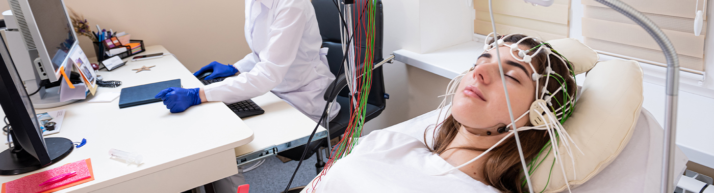
An EEG (electroencephalogram) records the brain's electrical activity through electrodes placed on the scalp. The EEG test mainly helps evaluate seizures and epilepsy, unexplained loss of consciousness, encephalopathy and some sleep disorders. An EEG does not image brain structure, and a normal EEG does not exclude epilepsy.
Түлхүүрийг авах
- Function, not structure: The EEG test shows electrical patterns; MRI and CT show anatomy. They answer different questions.
- A normal EEG is reassuring but not conclusive: Many people with epilepsy have a normal routine EEG between seizures.
- Abnormal does not automatically mean epilepsy: Some EEG patterns are benign variants or non-specific slowing.
- Safe and painless for most people: Risk comes mainly from activation procedures such as hyperventilation and photic stimulation, not from the recording itself.
- Preparation differs between units: Sleep-deprived, prolonged and video-EEG studies have different instructions. Your unit's written instructions take precedence.
Алсдаа
| Барааны | дэлгэрэнгүй |
|---|---|
| Юу хэмждэг | Electrical activity generated by cortical neurons, recorded from scalp electrodes |
| Нийтлэг заалтууд | Suspected seizures, known epilepsy, altered consciousness, encephalopathy, some sleep disorders, brain monitoring in ICU or theatre |
| Typical duration (routine EEG) | About 20-40 minutes of recording, plus roughly 15-30 minutes for electrode setup and removal |
| Longer studies | Sleep-deprived EEG, prolonged ambulatory EEG and inpatient video-EEG telemetry run from several hours to several days |
| Инвазив байдал | Non-invasive for scalp EEG; no needles or radiation |
| Pain | Usually painless; mild scratching or pressure during electrode application |
| Хэн гүйцэтгэж, тайлагнадаг вэ | EEG technologist records; a neurologist or neurophysiologist interprets |
| Гол хязгаарлалт | Normal EEG does not exclude epilepsy; EEG cannot show tumours, bleeds or strokes |
Мөн түүнчлэн
- Electroencephalogram, electroencephalography
- Тархины долгионы тест
- EEG brain test (commonly asked for as "brain ka EEG")
- Routine EEG, awake-sleep EEG, sleep-deprived EEG
- Video-EEG, EEG telemetry, long-term monitoring (LTM)
- cEEG (continuous EEG) in intensive care
What an EEG test is
Neurons in the brain communicate using small electrical currents. When large groups of neurons fire in a coordinated way, the summed voltage change can be detected at the scalp. An EEG amplifies and records these tiny voltages through electrodes fixed to the head, usually according to the international 10-20 system of electrode placement.
The output is a continuous trace of waveforms. A neurologist reads the background rhythm, its symmetry and reactivity, and looks for interictal epileptiform discharges (spikes, sharp waves, spike-and-wave complexes), focal or generalised slowing, and any electrographic seizure captured during the recording.
EEG is a functional test with excellent time resolution, it can show changes happening millisecond by millisecond, but poor spatial resolution. It is best at telling you хэрхэн the brain is behaving electrically, not ямар the brain looks like.
Why an EEG test is done
- Suspected epilepsy: To support a clinical diagnosis after one or more suspected seizures, and to help classify the seizure type and epilepsy syndrome (for example generalised versus focal epilepsy), which guides treatment choice.
- Known epilepsy: To assess seizure control, characterise absence seizures, and assist decisions about treatment.
- Events of uncertain nature: To help distinguish epileptic seizures from syncope, functional (dissociative) seizures, sleep phenomena or movement disorders, usually needing video-EEG.
- Өөрчлөгдсөн ухамсар эсвэл төөрөгдөл: To look for non-convulsive status epilepticus or the diffuse slowing of metabolic, toxic or infective encephalopathy.
- Encephalitis and specific patterns: For example, temporal periodic patterns that may accompany herpes simplex encephalitis, or characteristic patterns in some autoimmune encephalitides.
- Intensive care monitoring: Continuous EEG in patients with refractory seizures, after cardiac arrest, or during sedation titration.
- Intraoperative and pre-surgical work: Monitoring during carotid or neurosurgical procedures, and localising the seizure-onset zone before epilepsy surgery.
- Sleep medicine: EEG channels form part of polysomnography, used to stage sleep and evaluate narcolepsy, parasomnias and sleep-related epilepsy.
- Brain death evaluation: As an ancillary test where protocol requires it, within a strictly defined legal and clinical framework.
Энэ шинжилгээг хэн өгөх ёстой вэ
- Anyone who has had a first unprovoked seizure, once medically stabilised.
- People with recurrent blackouts, staring spells, unexplained falls or brief unresponsiveness where a seizure is a possibility.
- Children with staring episodes, unexplained developmental regression, or suspected absence or infantile epilepsy syndromes.
- Patients with epilepsy whose seizure pattern has changed or whose diagnosis is in doubt.
- Patients in ICU with unexplained prolonged coma or fluctuating consciousness after a convulsion.
- Эпилепсийн мэс засалд орохын тулд үзлэгт хамрагдаж буй хүмүүс.
Who usually does not need an EEG
- Uncomplicated headache or migraine, EEG is not a useful test for headache diagnosis.
- Typical vasovagal fainting with clear triggers and rapid full recovery, cardiac assessment is usually more relevant.
- Dizziness, giddiness or generalised weakness without features suggesting seizures.
- Routine "brain check-up" in a healthy person. EEG is not a screening test, and incidental benign variants can cause unnecessary anxiety and treatment.
- Diagnosing ADHD, anxiety, depression or schizophrenia. Despite what some clinics advertise, EEG-based "brain mapping" or qEEG is not an accepted diagnostic test for these conditions.
ЭЭГ юуг илрүүлж эсвэл үгүйсгэж чадахгүй вэ
This is the part most often missed in patient information, and it matters.
- EEG cannot exclude epilepsy. A single routine interictal EEG is normal in a substantial proportion of people who do have epilepsy. Repeat recordings, sleep-deprived studies or prolonged monitoring raise the yield, but a normal EEG never rules the diagnosis out. Epilepsy remains a clinical diagnosis.
- EEG does not image the brain. It cannot show a tumour, stroke, haemorrhage, abscess, neurocysticercosis, tuberculoma, hydrocephalus, demyelination or cortical malformation. Those need CT or MRI. An EEG may show slowing near a lesion, but it cannot tell you what the lesion is.
- EEG mostly sees the cortical surface. Activity arising deep in the brain, mesial temporal, orbitofrontal, insular or interhemispheric regions, may not reach scalp electrodes. Deep-seated seizure activity can be electrically silent on the surface.
- An abnormal EEG does not by itself diagnose epilepsy. Non-specific slowing has many causes, including drowsiness, drugs, fever and metabolic upset. Some sharp-looking patterns are benign normal variants seen in people who will never have a seizure.
- EEG cannot measure intelligence, personality, stress levels or "brain power". Nor can it detect Alzheimer's disease, autism or dyslexia.
- EEG cannot predict when the next seizure will occur or confirm that a person is safe to drive or swim.
- Standard EEG does not detect subtle peripheral or spinal problems: nerve and muscle problems need nerve conduction studies and EMG, which are entirely different tests.
If a result is reported as normal and your symptoms continue, that finding describes what was recorded during those minutes. It does not describe what happens at other times. Go back to the doctor who requested the test.
Холбоотой боловч өөр өөр шалгалтууд
| туршилтын | Асуулт нь хариулдаг | How it differs from a routine EEG |
|---|---|---|
| Routine (standard) EEG | Are there interictal epileptiform discharges or background abnormalities right now? | Short recording, usually awake with brief drowsiness; lowest sensitivity of the EEG family |
| Нойр дутуу EEG | Do abnormalities appear when the patient is sleep-deprived and falls asleep? | Patient sleeps less the night before; drowsiness and sleep increase the chance of capturing discharges. Not suitable for everyone, sleep deprivation can provoke seizures |
| Prolonged / ambulatory EEG | Do abnormalities or events occur across a day or more of ordinary life? | Recorded over 24 hours or longer, often at home; no continuous video in many setups, so event correlation is weaker |
| Video-EEG telemetry | What exactly happens electrically during the patient's typical attack? | Inpatient admission over days, synchronised video and EEG; the reference test for separating epileptic from non-epileptic events |
| Continuous ICU EEG (cEEG) | Is there non-convulsive seizure activity in a critically ill or comatose patient? | Performed at the bedside, often for days, with frequent review |
| Полисомнографи (нойрны судалгаа) | What is the sleep architecture, breathing and oxygenation overnight? | EEG is only a few channels among airflow, oximetry, ECG, effort belts and limb sensors; diagnoses obstructive sleep apnoea, which EEG alone cannot |
| Тархины MRI (эпилепсийн протокол) | Is there a structural cause such as hippocampal sclerosis or cortical dysplasia? | Anatomical imaging; complements EEG and does not replace it. Both are often done |
| Тархины компьютер томографи | Is there acute bleeding, a large lesion or hydrocephalus? | Fast, uses ionising radiation; used in emergencies. Shows structure only |
| NCS / EMG | Are the peripheral nerves and muscles working normally? | Tests nerves and muscles, not the brain. Commonly confused with EEG when booking |
| Evoked potentials (VEP, BAEP, SSEP) | Are specific sensory pathways conducting normally? | Measures the response to a defined stimulus rather than spontaneous brain activity |
| "Brain mapping" / qEEG | Marketed for psychiatric and learning conditions | Quantitative analysis of EEG data; not an accepted diagnostic test for ADHD, autism or psychiatric illness. Treat such claims with caution |
Which test comes first depends on the clinical problem. After a first convulsive seizure in an adult, urgent CT may come first if there was head injury or a focal deficit; EEG and MRI usually follow. For staring spells in a child, an EEG with hyperventilation is often the first and most informative test. For blackouts with palpitations, cardiac tests may take priority over EEG.
EEG-д хэрхэн бэлдэх вэ
Preparation instructions vary between hospitals and between EEG types. The written instructions from the unit performing your test take precedence over any general advice, including this page.
- Үс: Wash hair the evening before or on the morning of the test and let it dry. Do not apply oil, gel, serum, spray, wax or conditioner. Hair oil is a very common reason for poor electrode contact and repeat appointments. Remove hair extensions, tight braids or heavy clips if possible.
- Хоол хүнс: Most units do not require fasting, and a low blood sugar level can itself alter the EEG. Eat a normal meal unless your unit tells you otherwise.
- Кофейн ба өдөөгч бодисууд: Many units ask you to avoid tea, coffee, cola and energy drinks for several hours before the recording. Follow the interval your unit specifies rather than a fixed number from the internet.
- Эмийн эмчилгээ: Do not stop, reduce or delay any prescribed medicine, especially anti-seizure medication, on your own. Stopping anti-seizure medicine abruptly can trigger prolonged seizures. If a medication change is needed for the test, only the prescribing doctor should direct it, and usually only for planned inpatient monitoring.
- Bring a list: Current medicines and doses, a description of your episodes, their frequency, triggers and duration, and previous EEG or MRI reports. A phone video of a typical event recorded by a family member is often extremely helpful to the neurologist.
- Компани: Bring a relative or friend, particularly if you have frequent seizures, are sleep-deprived for the test, or may feel drowsy afterwards.
- Хувцас: Loose, comfortable clothes with an open neckline.
Нойр дутуу EEG
If a sleep-deprived study is requested, the unit will tell you how much sleep to restrict. Do not drive yourself to or from the appointment. Sleep deprivation can provoke seizures in susceptible people, so arrange supervision for the whole day. Tell the unit in advance if you have poorly controlled epilepsy, severe mental health illness such as bipolar disorder, obstructive sleep apnoea, or work night shifts.
Хүүхэд
Paediatric EEGs are often arranged around the child's natural nap time. Restricting a young child's sleep should only be done if the unit specifically asks and the parents can supervise safely. Bring a favourite toy, feed, pillow or blanket. Explaining beforehand that the electrodes are like stickers that "listen" and do not give shocks reduces distress substantially. Sedation is used selectively and only under supervision, as sedation itself changes the EEG.
Чихрийн шижин өвчний үед глюкоз бууруулах эмчилгээ хийлгэж байна
If you take insulin or sulfonylureas, do not skip meals for an EEG. Hypoglycaemia is dangerous and also distorts the recording. Tell the unit at booking and bring a fast-acting sugar source.
Жирэмсний
Scalp EEG uses no radiation and no contrast, and is performed during pregnancy where clinically indicated. Hyperventilation and photic stimulation may be modified or omitted, tell the technologist you are pregnant.
Older adults, CKD, heart failure and reduced mobility
Tell the unit about any difficulty lying flat, breathlessness, or the need to keep to a fluid restriction. Positioning can be adapted. Metabolic disturbance in advanced kidney disease and some medications commonly cause diffuse slowing on the trace, and the reporting neurologist needs to know about these to interpret the record properly.
EEG-ийн үед юу болдог вэ
- Хэмжилт ба тэмдэглэгээ: The technologist measures your head and marks electrode positions. Sites are lightly cleaned with a mildly abrasive gel, you may feel scratching or rubbing.
- Электродын хэрэглээ: Around 21 or more small cup electrodes are fixed with conductive paste, or a cap is fitted. This takes roughly 15-25 minutes.
- Суурь бичлэг: You lie or sit still with your eyes closed, opening and closing them when asked.
- Гипервентиляци: You may be asked to breathe deeply for around three minutes. Light-headedness and tingling in the hands are common and settle quickly. This step is omitted in people with recent stroke, significant heart or lung disease, sickle cell disease, moyamoya disease, or recent intracranial bleeding.
- Photic stimulation: A lamp flashes at varying frequencies with eyes open and closed, to look for a photoparoxysmal response. It is stopped immediately if abnormal activity appears.
- Унтах: Drowsiness and sleep are actively encouraged, since many discharges appear only in light sleep.
- Дараа нь: Electrodes are removed and the paste wiped off. Some residue may remain in your hair until you wash it. There are no restrictions after a standard EEG, unless you were sleep-deprived, sedated or had a seizure during recording.
Процесс хэр удаан үргэлжлэх вэ
- Ердийн EEG: about 45-75 minutes in the department in total, of which 20-40 minutes is actual recording.
- Sleep-deprived or awake-and-sleep EEG: roughly 1-3 hours, sometimes longer if sleep is slow to come.
- Амбулаторийн EEG: setup takes under an hour, then 24-72 hours of recording at home.
- Video-EEG telemetry: an inpatient stay typically lasting several days, sometimes longer, until enough typical events are captured.
- Neonatal and paediatric EEG: variable, since recordings are timed around feeding and sleep.
Reporting is separate from recording. The trace must be reviewed page by page by a neurologist or neurophysiologist, so the report is not available immediately after the test. Ask the unit when to expect it.
Таны EEG тайланг ойлгох
An EEG report is descriptive prose, not a number with a reference range. There is no "EEG normal value" in the way there is for haemoglobin or blood sugar. A typical report comments on:
- Background rhythm: The dominant posterior rhythm in a relaxed adult with eyes closed is usually in the alpha range and attenuates on eye opening. The expected frequency is age-dependent, infants and young children normally have slower backgrounds, which is not a sign of disease.
- Symmetry and organisation: Whether the two hemispheres look comparable.
- Реактив байдал: Whether the trace changes appropriately with eye opening, sound or stimulation.
- Sleep architecture: Whether normal drowsiness and sleep features were recorded.
- Epileptiform abnormalities: Spikes, sharp waves, spike-and-slow-wave complexes; whether focal or generalised.
- Удаашрах: Focal slowing may suggest a localised problem; generalised slowing suggests a diffuse process such as encephalopathy, sedation or sleep.
- Response to activation: Findings on hyperventilation and photic stimulation.
- Artefacts: Muscle tension, eye movement, sweating, chewing, ECG or mains interference.
About "brain wave types"
Delta, theta, alpha, beta and gamma are simply frequency bands used to describe the trace. They are not personality traits or health scores. A band is only meaningful in context: theta and delta are entirely normal in sleep and in young children, and prominent beta is commonly produced by benzodiazepines and some other medicines rather than by anxiety. Claims that EEG frequency bands can diagnose stress, ADHD, depression or "low concentration" are not supported by mainstream neurology, and the popular "significance" lists circulated online overstate what these bands mean.
Normal, abnormal and in between
- Normal EEG: No epileptiform discharges or significant slowing were seen during that recording. This does not exclude epilepsy or any other brain disorder.
- Abnormal EEG: Names a specific finding. The meaning depends entirely on your symptoms, examination, imaging and history.
- Benign variants: Several sharply contoured patterns look alarming but have no clinical significance. Experienced readers identify them precisely so that people are not labelled with epilepsy wrongly.
An EEG report is never a diagnosis on its own. Only the doctor who requested the test can interpret it together with your clinical picture.
ЭЭГ-ийн үр дүн төөрөгдүүлж болох үед
Худал хэвийн бус харагдаж болох олдворууд
- Drowsiness and normal sleep: Slow activity in drowsiness can be mistaken by inexperienced readers for pathological slowing.
- Эмийн нөлөө: Benzodiazepines, barbiturates and some other drugs produce prominent fast activity; sedatives and opioids produce slowing.
- Metabolic and systemic upset: Uraemia, liver failure, low sodium, hypoglycaemia, hypothyroidism, sepsis and fever all cause diffuse slowing that is not a primary brain disease.
- Benign normal variants: Sharp-looking patterns that mimic epileptiform discharges.
- Artefacts: Eye flutter, chewing, tremor, scalp sweating, a loose electrode, mobile phones or nearby equipment can all mimic abnormalities.
- Гипервентиляци: Marked slowing during deep breathing is a normal response in children and young adults, especially if blood sugar is low.
- Саяхан таталт өгсөн: Post-ictal slowing may persist for hours to days and can be over-interpreted.
Худал тайвшруулж болох олдворууд
- Short recordings: A 20-minute sample of a 24-hour brain may simply miss intermittent discharges.
- Anti-seizure medication: Treatment can suppress interictal discharges, so a normal EEG in someone already on treatment says little.
- Deep sources: Mesial temporal, orbitofrontal and insular activity often does not reach scalp electrodes.
- No sleep achieved: If the person stayed fully awake throughout, the yield drops, since many discharges appear in light sleep.
- Activation not performed: If hyperventilation or photic stimulation was omitted for safety, certain responses cannot be assessed.
- цаг хугацаа: An EEG done long after the event is less informative than one done within 24-48 hours of a first seizure.
Эрсдэл ба аюулгүй байдал
Different types of EEG carry different risk profiles. It is not accurate to describe the whole family with one safety statement.
Хуйхны ердийн EEG
Very low risk. No radiation, no contrast, no needles. Possible minor issues include scalp irritation or redness from the abrasive gel, an uncommon allergic reaction to collodion or paste, discomfort during electrode removal, and residual paste in the hair. Collodion, used in some units for longer recordings, has a strong odour and requires an acetone-based remover.
Идэвхжүүлэх журам
Hyperventilation can cause light-headedness, tingling and, rarely, a seizure, that is partly its purpose. It is avoided in people with significant cardiac or respiratory disease, recent stroke or intracranial haemorrhage, sickle cell disease and moyamoya disease. Photic stimulation may provoke a seizure in photosensitive individuals; recordings are supervised and stimulation is stopped as soon as a photoparoxysmal response appears.
Нойр дутуу EEG
Sleep deprivation increases seizure risk in susceptible people and can worsen mood in some psychiatric conditions. Driving after a sleep-deprived study is unsafe.
Prolonged video-EEG with medication reduction
Carried out as an inpatient precisely because it carries real risk. Deliberately reducing anti-seizure medication to capture seizures can lead to seizure clusters, status epilepticus, injury from falls or post-ictal confusion. Continuous nursing observation and an emergency plan are essential. This is never done at home or self-directed.
Invasive EEG
Intracranial EEG with subdural or depth electrodes is a neurosurgical procedure used only in selected epilepsy-surgery candidates. It carries surgical risks including bleeding and infection, and is entirely different from a scalp EEG.
Үр дүн болон эрсдэл нь тухайн эмнэлзүйн нөхцөл байдлаас хамааран өөр өөр байдаг.
Аюултай тугнууд: хэзээ тусламж хүсэх вэ
Emergency now (call an ambulance or go to the nearest emergency department)
- A convulsion lasting more than five minutes, or repeated seizures without full recovery in between.
- Seizure followed by persistent unconsciousness, confusion that does not clear, or difficulty breathing.
- A first-ever seizure in an adult.
- Seizure with head injury, significant bleeding, suspected drowning or aspiration.
- Seizure in pregnancy, or with fever and neck stiffness or a new severe headache.
- New weakness on one side, facial droop or difficulty speaking after an event.
- A seizure in a person with diabetes who may be hypoglycaemic.
Same day (contact your doctor or neurology service today)
- A clear increase in seizure frequency or a change in seizure pattern in known epilepsy.
- Missed doses of anti-seizure medicine, or vomiting that prevents taking medicine.
- New rash, mouth ulcers or fever after starting an anti-seizure medicine.
- Seizure occurring while recovering from a sleep-deprived EEG.
- Brief episodes of confusion or unresponsiveness in an older adult that are new.
Ердийн цаг товлох
- Discussing an EEG report and what it means for your diagnosis and treatment.
- Symptoms continuing despite a normal EEG, return to the requesting doctor for reassessment.
- Side effects that are troublesome but not alarming.
- Questions about driving, work, schooling, contraception or pregnancy planning with epilepsy.
Онцгой нөхцөл байдал
Жирэмсний
EEG is safe in pregnancy and is often used when new seizures occur or existing epilepsy changes. Late-pregnancy seizures also raise the possibility of eclampsia, which is an obstetric emergency assessed clinically and biochemically, not by EEG. Never alter anti-seizure medication during pregnancy without the treating neurologist and obstetrician.
Нярай болон нярай хүүхдүүд
Neonatal seizures are frequently subtle or entirely electrographic, so EEG or amplitude-integrated EEG monitoring is central to neonatal care. Neonatal EEG has its own maturational patterns and requires specialist reporting; adult criteria do not apply.
Хүүхэд ба өсвөр насныхан
Children with absence epilepsy often show a characteristic generalised discharge pattern during hyperventilation, making EEG particularly valuable. Photosensitivity is more common in adolescents. Some childhood epilepsy syndromes show discharges that markedly increase in sleep, which is why sleep recordings are frequently requested.
Хуучин насанд хүрэгчид
Late-onset seizures are commonly related to previous stroke or other structural change, so imaging is essential alongside EEG. Non-convulsive status epilepticus is an under-recognised cause of prolonged confusion in older people, and EEG is the only way to identify it.
Critically ill and comatose patients
Continuous EEG detects electrographic seizures that produce no visible movement, particularly after convulsive status epilepticus, cardiac arrest, head injury or stroke.
People with cardiac devices or metal implants
Unlike MRI, EEG has no magnetic field and pacemakers, ICDs and metallic implants do not preclude the recording. Tell the technologist about any implant, as a deep brain stimulator or vagus nerve stimulator produces recognisable interference that the reader should know about.
Scalp conditions and head wounds
Open wounds, surgical sites, severe eczema or active scalp infection may require electrodes to be repositioned or the test postponed.
Энэтхэгийн нөхцөлд EEG
- Epilepsy is common and often untreated. India carries a large share of the global epilepsy burden, and a substantial treatment gap persists, particularly in rural areas, where access to a neurologist and to EEG services is limited. Many people are diagnosed and treated without ever having an EEG, and some are treated for epilepsy they do not have.
- Neurocysticercosis and tuberculomas are among the leading identifiable causes of new-onset seizures in many parts of India. These are structural diagnoses made on CT or MRI, an EEG cannot detect them. Anyone with new seizures should have appropriate neuroimaging as well as EEG.
- Хүүхдийн халууралт таталт are common. Simple febrile seizures generally do not require an EEG; the immediate priority is identifying the cause of fever and excluding central nervous system infection.
- Hair oil and long hair are a genuine practical issue. Oiled hair prevents good electrode contact and is a frequent cause of repeat appointments. Wash the hair and keep it free of any product before your appointment.
- Гутаан доромжлол ба буруу мэдээлэл. Epilepsy still carries stigma in many communities, and families sometimes delay medical care in favour of unproven remedies. Under the Drugs and Magic Remedies (Objectionable Advertisements) Act 1954, advertisements claiming to cure epilepsy are prohibited. Be cautious about any clinic promising a guaranteed cure, "brain mapping"-based diagnosis of learning or behavioural problems, or EEG-based treatment for psychiatric illness.
- Жолоо барьж байна. Fitness to drive after seizures is governed by medical advice and motor vehicle regulations. Discuss this openly with your neurologist rather than relying on informal advice.
- Мэдээллийн хамгаалалт. EEG traces, videos and reports are sensitive personal health data. Video-EEG in particular records you continuously. Ask how footage is stored, who may access it, and how consent is obtained and withdrawn, as required under the Digital Personal Data Protection Act 2023.
Энэтхэг дэх зардал ба даатгал
EEG charges differ widely between government hospitals, trust hospitals, standalone neurodiagnostic centres and corporate hospitals, and between cities. A routine outpatient EEG is generally one of the less expensive neurological investigations, while prolonged video-EEG telemetry involves an inpatient admission and costs considerably more. Always ask for a written estimate before booking.
Зардлыг юу өдөөдөг вэ
- Судалгааны төрөл: routine EEG versus sleep-deprived, ambulatory, or multi-day video-EEG telemetry.
- Duration of recording and whether an inpatient bed, nursing observation and continuous video are required.
- Сувгуудын тоо and whether high-density montages are used.
- Sedation or anaesthetic support, occasionally needed for young children or uncooperative patients.
- Тайлагнах: neurologist versus subspecialist epileptologist review; urgent or after-hours reporting.
- Тохиргоо: outpatient department, bedside ICU recording, or intraoperative monitoring.
- Нэмэлтүүд: repeat studies, MRI epilepsy protocol and blood









 Ченнай хотын ойролцоох хамгийн сайн эмнэлэг


