


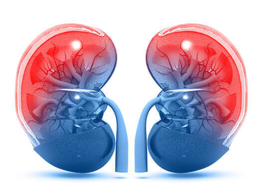

.png)


- Imaging
- Laboratory
- Ultrasound: Ultrasound images are created using sound waves and are able to gather real-time images of both anatomy as well as function. It is a non-invasive scanning process that does not utilize any ionizing radiation that could be harmful to patients. It helps us in the initial evaluation of kidneys to look for any solid tumor.
- CT SCAN: It is a diagnostic imaging procedure that uses x-rays to build cross-sectional images of the kidney. We inject intravenous contrast to further characterize the kidney tumor and to look for its extent.
- PET-CT: It is a tool used to provide more information in diagnosing primary kidney cancer and to look for any metastases to other organs, which helps in further management.
- MRI: A tool that uses the interaction of radio waves and magnetic field, which is processed in a high-speed computer system to produce scan pictures and aids in a detailed, informative study about the kidney tumor, its blood supply, location and its extent. Special sequences like contrast study (Special dye injected through the veins) and diffusion helps to further characterize it.
- Urine cytology: Renal cell cancer will have blood in their urine. If the patient has transitional cell carcinoma (in the renal pelvis, the ureter, or the bladder), sometimes a special test of the urine sample (called urine cytology) will show actual cancer cells in the urine.