


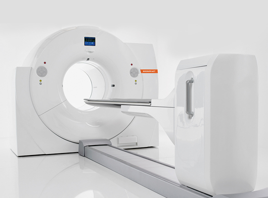
Positron Emission Tomography - PET-CT Scan
Introduction
The scan uses a special dye containing radioactive tracers (Isotope labelled Glucose molecules). These tracers are either swallowed, inhaled, or injected into a vein in the arm depending on what part of the body is being examined. Certain organs and tissues then absorb the tracer.
When detected by a PET scanner, the tracers let the doctor see how well the organs and tissues are working.
The tracer will collect in areas of higher chemical activity, which is helpful because certain tissues of the body, and certain diseases, have a higher level of chemical activity. These areas of disease will show up as bright spots on the PET scan.
Features
PET-CT combines the fine structural detail of CT with PET’s ability to detect changes in cell function. This combination allows for earlier and more accurate detection of disease than either CT or PET alone
Indications and procedures
Most commonly used to detect:Cancer, heart problems, brain disorders PET scans are useful both for detecting cancer and for:
- diagnosing if the cancer has spread
- observing if a cancer treatment is working
- checking for a cancer recurrence
- Read More
Advantages
PET/CT images supplement the information obtained from conventional studies such as CT, MRI, and Ultrasound. It also helps in accurate diagnoses, develop more targeted treatment plans, and do better, less-invasive treatment monitoring, which should result in improved patient outcomes.