


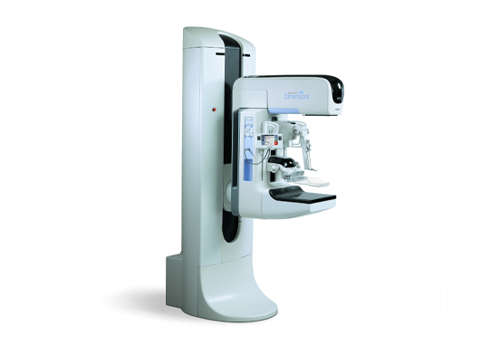
Contrast-Enhanced Mammogram Technology
Introduction
Contrast-enhanced spectral mammography (CEM) is a specific type of mammogram exam that uses medical imaging contrast to create a special picture of the breast tissue.
Essentially, the image that CEM creates is a composite of two images taken simultaneously.
Indications
- Evaluation/ Problem Solving – difficult to interpret mammograms
- Identify potential undetected malignancies
- Evaluate the extent of disease pre-operatively
- Assessment of response to chemotherapy
- Patients contraindicated for MRI, e.g. claustrophobia or pacemakers
Procedures
A technician will start an IV and begin administering the contrast intravenously. It takes two minutes for the contrast to spread throughout the body following which, the technologist will take images of both of the breasts.
Contrast-enhanced spectral mammography (CEM) only takes a little longer than a standard mammogram.
Advantages
- Able to identify breast cancer before it is visible on a standard mammogram.
- A useful alternative to MRI or where MRI is contraindicated (e.g. gadolinium allergy or claustrophobia) for staging, or follow-up post-treatment.
