


.png)


- Imaging
- Laboratory
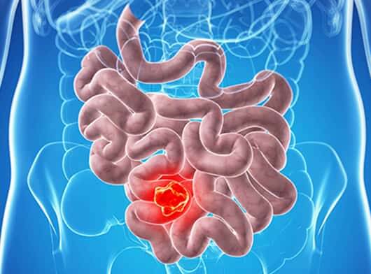
- CT scan is a diagnostic imaging procedure that uses x-rays to build cross-sectional images of the body. It also helps to identify the spread of cancer. The doctor may perform a CT scan of the abdomen to help identify the tumor and ascertain the degree of spread.
- PET CT provides functional and morphological details by utilizing radiation derived from Isotope labeled Glucose molecules to detect cellular glucose uptake in cancer. PET CT is done in Jejunal cancers to assess spread to regional nodes or distant metastases to other parts of the body.
- Endoscopy/ Enteroscopy - is done in some cases to visualize and biopsy the tumor. It is a procedure that uses a thin, flexible tube, attached with a camera, called an enteroscope, to examine the interior of a small bowel. It may be used to collect a tissue sample for biopsy. However, not all cases of Jejunal cancers require this procedure.
-
Biopsy It is the removal of a small amount of tissue for further pathology examination and to detect the type of tumor. A Biopsy can be CT/ Ultrasound/ Endoscope guided based on the location of the lesion.
A Biopsy may not be required for Jejunal tumors where the lesion is surgically resectable. However, if a physician wants to determine the type of tumor, a biopsy might be ordered.
- Pathology - The tissue sample taken is sent for further evaluation like Histopathological examination (HPE) and Immunohistochemistry (IHC) to determine the type of disease and its grading.