


.png)


- Imaging
- Laboratory
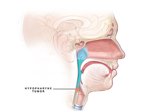
- Fiber optic hypopharyngoscopy - The doctor examines the hypopharynx with the help of small mirrors or fiber optic endoscope by passing the thin endoscope through the nose into the pharynx. This is done under local anesthesia.
- Contrast-enhanced CT scan of the neck - can confirm the presence of a tumor and also provides detailed information regarding the abnormality, its location, size, relationship to other structures, and tissue characteristics, particularly involvement/invasion or compression of surrounding structures. This is critical to treatment planning.
- Positron emission tomography (PET) scan - Positron emission tomography (PET) scan does functional and morphological detail scanning by utilizing radiation derived from isotope labeled glucose molecules that enable detection of cellular glucose uptake in cancer.
- MRI - Magnetic resonance imaging (MRI) uses the interaction of radio waves and magnetic field, which will be processed in a high-speed computer system to produce detailed scan pictures of the tissue, organs, bones, ligament and cartilage.
- Biopsy – This is done with the help of a fiber optic endoscope which is passed through the nose into the pharynx; the doctor collects a sample of tissue for further evaluation.
- Pathology – Sample collected is sent for further investigations such as Histopathological evaluation (HPE) and Immunohistochemistry (IHC) to determine the type of cancer and grading.