


.png)


- Medical history and physical exam
- Blood Tests
- Lymph node biopsy
- Imaging tests
- On suspecting a leukemia, your doctor will want to take a complete medical history to check for symptoms and possible risk factors. You'll also be asked about your family medical history and your general health. A physical exam will be done to look for possible signs of leukemia and other health problems. During the exam, your doctor will pay close attention to your lymph nodes, abdomen (belly), and other areas that might be affected. Your doctor may also order tests to check your blood cell counts. If the results suggest leukemia, you may be referred to a hematologist, a doctor who specializes in treating blood disorders (including blood cancers like leukemia).
- Tests used to diagnose and classify leukemia:
- Tests will need to be done on your blood and bone marrow to be certain of a leukemia diagnosis. Other tissue and cell samples may also be needed to help guide treatment.
- Blood samples for tests for CLL will be taken from a vein in your arm. Many different tests are done.
- Complete blood count and blood cell exam (Peripheral Blood Smear) :
- The complete blood count or CBC measures the different cells in your blood, such as the red blood cells, the white blood cells, and the platelets. This test is often done along with a differential (or diff) which looks at the numbers of the different types of white blood cells. These tests are often the first ones done when a blood problem is suspected.
- People with CLL have too many lymphocytes. (This may be called lymphocytosis.) Having more than 10,000 lymphocytes/mm³ (per cubic millimeter) of blood strongly suggests CLL, but other tests are needed to know for sure. You might also have low levels of red blood cells and platelets.
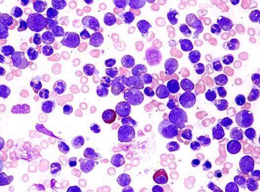
- A sample of blood is looked at under the microscope (called a peripheral blood smear). If you have CLL, the blood smear could show many abnormal looking lymphocytes called smudge cells.
- Flow cytometry
- Other blood tests
- Bone marrow tests
- Routine microscopic exams
- Gene tests
- Cytogenetics
- Fluorescent in situ hybridization (FISH)
- Molecular tests
This test is important in diagnosing CLL. It uses a machine that looks for certain substances (markers) on or in cells that help identify what types of cells they are.
This test can be used to see if the lymphocytes in a sample of blood contain CLL cells. Flow cytometry can also be used to look for CLL cells in bone marrow or other fluids.
Your blood immunoglobulin (antibody) levels may be tested to check if you have enough antibodies to fight infections, especially if you've recently had many infections. Another blood protein called beta-2-microglobulin may be measured. High levels of this protein generally mean a more advanced CLL.
Bone marrow aspiration and biopsy are done to get bone marrow samples for testing. They're usually done together. The samples are usually taken from the back of the pelvic (hip) bone.
A pathologist (a doctor specializing in lab tests) looks at the bone marrow samples under a microscope. They may also be reviewed by your hematologist/oncologist (a doctor specializing in blood diseases and cancer).
Stains and/or antibody tests such as cytochemistry, immunocytochemistry, immunohistochemistry, and flow cytometry may be used on the bone marrow samples to diagnose CLL. You can learn more about these tests at Tests Used on Biopsy and Cytology Specimens to Diagnose Cancer.
For this test, bone marrow cells (or sometimes cells from the blood or other tissues) are grown in the lab, then their chromosomes are examined under a microscope. Because it takes time for the cells to start dividing, this test usually takes weeks to complete.
This information may be helpful to determine a patient's prognosis (outlook), but it needs to be looked at along with other factors, such as the stage of CLL. The loss of part of chromosome 13 is usually linked with a slower-growing disease and a better outlook, while defects in chromosomes 11 or 17 often indicate a poorer outlook. Trisomy 12 doesn't seem to have much of an effect on prognosis. Deletion 17p abnormality is associated with a high risk C.L.L.
This chromosome test can be used to look at the cells’ chromosomes and DNA. It uses special fluorescent dyes that only attach to specific parts of particular chromosomes. FISH is used to look for certain genes or chromosome changes (not just any change). It can be used on regular blood or bone marrow samples, too.
Immunoglobulins, the antibodies that help your body fight infections, are made up of light chains and heavy chains. Whether the gene for the immunoglobulin heavy chain variable region (IGHV or IgVH) has changed (mutated) can help your doctor know how aggressive your CLL is. That gene is looked at in a test called cDNA sequencing.
-
In a lymph node biopsy, all or part of a lymph node is removed so it can be examined under the microscope to see if it contains cancer cells. This is often done to diagnose lymphomas, but only rarely needed for CLL. It may be done if a lymph node has grown very large and the doctor wants to know if the leukemia has changed (transformed) into a more aggressive lymphoma.
-
Imaging tests use x-rays, sound waves, or magnetic fields (CT/ Ultrasound/ MRI) to create pictures of the inside of the body. Imaging tests are not done to diagnose CLL, but they may be done for other reasons, for instance to help find a suspicious area that might be cancer, to learn how far a cancer may have spread, or to help see if treatment is working.