


সরাসরি উত্তর
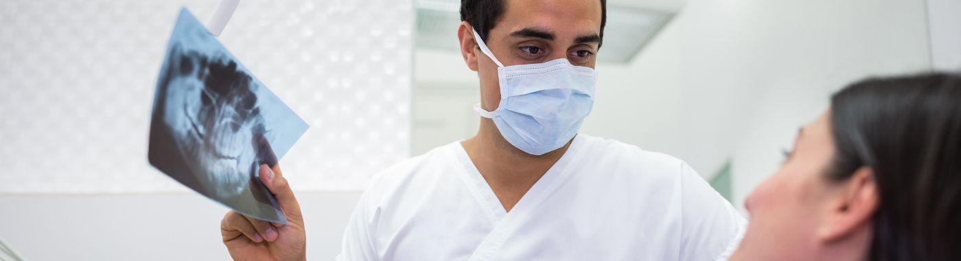
Dental X-rays are low-dose radiographs of the teeth, jaws and supporting bone. Dental X-rays show decay between teeth, bone loss, root infection, impacted teeth and jaw lesions that a visual check cannot reveal. Dental X-rays do not assess soft-tissue lesions or pain cause directly, and a normal film does not exclude disease.
কী টেকওয়েস
- Different views answer different questions: bitewing for decay between teeth, periapical for root and surrounding bone, panoramic (OPG) for a whole-jaw overview, CBCT for three-dimensional planning.
- Doses are low but not zero: radiographs are taken when a clinical question needs answering, not as a routine yearly ritual.
- Films show hard tissue: X-rays cannot reliably show gum inflammation, nerve vitality, early enamel demineralisation or soft-tissue cancers.
- ব্যাখ্যার জন্য প্রেক্ষাপট প্রয়োজন: images must be read alongside your symptoms and examination by the dentist who requested them.
- Tell the team if you are or may be pregnant so shielding and justification can be discussed before exposure.
এক পলকে
| বৈশিষ্ট্য | বিস্তারিত |
|---|---|
| এটি যা চিত্রিত করে | Teeth, roots, alveolar bone, jaws, some sinus and TMJ structures |
| সাধারণ প্রকার | Bitewing, periapical, occlusal, panoramic (OPG), lateral cephalogram, CBCT |
| রেডিয়েশন | Ionising, low dose; varies widely by view (intraoral lowest, CBCT highest) |
| ব্যথা | Painless; intraoral sensors may feel uncomfortable or trigger gagging |
| Time in the chair | A single intraoral film takes seconds; a full set or OPG usually a few minutes |
| Contrast or injection | ব্যবহার করা হয় না |
| কে এটা পড়ে | Dentist, oral and maxillofacial surgeon, or oral radiologist |
| ফলাফল | Digital images appear on screen almost immediately; formal reporting depends on the unit |
এই নামেও পরিচিত
- Dental radiograph, dental film
- ওপিজি / orthopantomogram / "full mouth X-ray" (panoramic)
- আইওপিএ: intraoral periapical, the term most used in Indian clinics
- আরভিজি: radiovisiography, the common Indian term for a digital intraoral sensor film
- সিবিসিটি / dental CT / 3D dental scan
- "Daant ka X-ray", "teeth ka scan"
What dental X-rays are
A dental X-ray is an image formed by passing a small, tightly collimated beam of X-rays through the jaw onto a sensor or film. Dense structures such as enamel, dentine, bone and metal fillings absorb more of the beam and appear pale; decay, infection, cysts and soft tissue absorb less and appear darker.
Most Indian clinics now use digital sensors (RVG) or phosphor plates rather than film. Digital systems generally allow a lower exposure per image and give an immediate on-screen picture, but image quality still depends on sensor placement, beam angle and patient movement.
Intraoral views
- Bitewing: shows the crowns and the contact areas of upper and lower back teeth together. The standard view for decay between teeth and for checking bone level at the crest.
- Periapical (IOPA): shows the whole tooth from crown to root tip plus surrounding bone. Used for toothache, root canal treatment, trauma and suspected abscess.
- Occlusal: a broader view of an arch, used mainly in children, for retained or unerupted teeth, and for some cysts or salivary stones in the floor of the mouth.
Extraoral views
- Panoramic (OPG): one curved image of both jaws. Good for an overview, wisdom teeth, jaw fractures, large lesions, pre-denture assessment. Poorer detail than intraoral films, with overlap in the front teeth and geometric distortion.
- Lateral cephalogram: a side view of skull and jaws used for orthodontic and jaw-surgery planning, not for decay.
- CBCT: a three-dimensional data set used for implant planning, complex root canal anatomy, impacted teeth near nerves, jaw pathology and some trauma. CBCT gives far more anatomical information than a plain film and also a higher radiation dose, so it is justified case by case rather than used as a screening tool.
Why dental X-rays are done
- Decay between and beneath teeth that is hidden from direct view, and decay under existing fillings or crowns.
- Periodontal assessment: the pattern and extent of bone loss around teeth.
- Root and periapical problems: abscess, cyst, root fracture, resorption, failed previous root canal treatment.
- Impacted or unerupted teeth, especially wisdom teeth and canines, and their relation to nerves and adjacent roots.
- চিকিত্সা পরিকল্পনা: root canal length, extraction difficulty, implant site bone volume, orthodontic assessment.
- ট্রমা: suspected tooth or jaw fracture, displaced teeth.
- আকস্মিক পর্যবেক্ষণ: jaw cysts, tumours, retained root fragments, sinus floor changes, calcified structures.
- শিশু: monitoring eruption sequence, missing or extra teeth, and decay risk in primary molars.
Who should consider a dental X-ray
- সাথে যে কেউ persistent toothache, sensitivity to biting, facial swelling or a bad taste from one area.
- মানুষের সাথে closely spaced back teeth or previous decay between teeth, where visual checks are unreliable.
- মানুষের সাথে bleeding gums, loose teeth or gum recession, to map bone loss.
- Anyone planned for root canal treatment, extraction, implant, crown, denture or orthodontics.
- People after facial or dental injury.
- Children with delayed eruption, crowding, or a high decay risk, on the dentist's judgement.
- লোকেরা bisphosphonates or other bone-modifying drugs, or after head and neck radiotherapy, before extractions.
Radiographs are not recommended simply because it has been a year since the last set. A dentist decides based on your examination, risk of decay, symptoms and planned treatment. Intervals differ widely between individuals, and results vary depending on individual clinical circumstances.
What dental X-rays cannot detect or exclude
- মুখের ক্যান্সার: dental X-rays are not a screening test for oral cancer. Most mouth cancers begin in soft tissue, tongue, buccal mucosa, palate, and are invisible on a radiograph until bone is involved. A normal X-ray says nothing about a mouth ulcer, white or red patch, or lump. These need direct clinical examination and, where indicated, biopsy. This matters in India, where tobacco- and areca nut-related oral cancer is common.
- মাড়ির প্রদাহ: gingivitis, pocket depth and bleeding are clinical measurements, not radiographic ones. X-rays show the bone that has already been lost, not current disease activity.
- Early enamel demineralisation and many early occlusal (biting-surface) cavities are frequently missed; roughly a third or more of mineral loss may be needed before decay becomes visible.
- Nerve vitality: an X-ray cannot tell whether a pulp is alive, dying or dead. That needs vitality testing and symptom history.
- ফাটা দাঁত সিন্ড্রোম: hairline crown or root fractures often do not appear on plain films.
- Cause of facial pain: sinus disease, temporomandibular joint pain, trigeminal neuralgia, neuropathic and referred pain are usually not diagnosed on dental films.
- Early peri-implant or periapical change: bone loss must reach a certain extent before it shows, and two-dimensional films collapse depth, hiding lesions behind dense roots.
- Soft-tissue extent of swelling or infection: spreading facial infection is judged clinically; deep spread may need CT.
A finding not identified on a dental X-ray is not the same as a finding excluded. If your pain, swelling or symptom continues despite a normal radiograph, return to your dentist, repeat imaging, a different view, vitality testing or referral may be needed.
সম্পর্কিত কিন্তু ভিন্ন পরীক্ষা
| পরীক্ষা | প্রশ্ন করলে উত্তর পাওয়া যায় | যেখানে এটি কম পড়ে |
|---|---|---|
| বাইটউইং | Is there decay between back teeth? Is crestal bone level normal? | Does not show root tips or periapical lesions |
| Periapical (IOPA) | What is happening at this tooth's root and surrounding bone? | Covers only two or three teeth; poor for decay contacts if angled badly |
| Panoramic (OPG) | Overview of both jaws, wisdom teeth, large lesions | Distortion, overlap, insufficient detail for early decay or fine bone changes |
| Lateral cephalogram | Jaw and facial skeletal relationships for orthodontics | No information on decay or root pathology |
| সিবিসিটি | Three-dimensional bone volume, nerve position, complex anatomy | Higher dose; poor soft-tissue contrast; metal artefacts; not a decay screening tool |
| Medical CT / MRI | Soft tissue, tumours, spreading infection, TMJ discs, salivary glands | Not used for routine dental decay or bone-level assessment |
| Ultrasound of face or neck | Soft-tissue swellings, lymph nodes, salivary glands | Cannot see inside teeth or through bone |
| Biopsy / cytology | Is a mucosal patch or ulcer malignant? | No imaging substitute; X-rays cannot answer this |
None of these views is universally superior. The right one depends on the clinical problem: a single aching molar usually needs an IOPA, not an OPG; implant planning usually needs CBCT, not a bitewing.
কিভাবে তৈরী করতে হবে
Preparation is minimal and there is no fasting. Protocols, positioning aids, shielding practice and image-sharing methods differ between clinics and hospitals, and your unit's written instructions take precedence over any general guidance here.
- Tell the team if you are pregnant or might be.
- Remove metal from the imaging field: earrings, nose pins, facial piercings, spectacles, hairpins, removable dentures, orthodontic appliances and hearing aids, if asked.
- Bring previous images and reports, including films on CD, USB or phone photographs, so change over time can be judged.
- Mention a strong gag reflex, jaw stiffness, limited mouth opening, or recent facial injury: alternative sensor sizes or extraoral views may be used.
- Mention past head and neck radiotherapy or bone-modifying medicines before extraction planning.
আপনি যদি গর্ভবতী হন বা বুকের দুধ খাওয়ান
Dental treatment need not be postponed for pregnancy, and untreated dental infection carries its own risks. Radiographs may still be taken during pregnancy when clinically justified, using the smallest number of exposures and appropriate shielding, ideally limited to what treatment requires. Discuss timing and necessity with your dentist and obstetrician. Breastfeeding is unaffected by dental X-rays.
If you are bringing a child
Children are more radiosensitive than adults, so exposures are kept to the minimum and paediatric settings and smaller sensors are used. Bring the child's previous films. A parent may be allowed to stay and support the child, wearing protection, depending on unit policy.
If you have limited mouth opening, tremor or cognitive difficulty
Holding an intraoral sensor still can be hard. Tell the team in advance; extraoral views such as an OPG may be substituted, or a holder and assistance arranged.
What happens during a dental X-ray
- পজিশনিং: for intraoral films you sit in the chair and a small sensor or plate is placed in your mouth, often in a plastic holder that you bite gently. For an OPG you stand or sit, bite on a plastic post, and the machine rotates around your head.
- প্রতিরক্ষা: protective aprons or thyroid collars may be offered depending on the view and current unit policy; modern collimation already restricts the beam to a small area.
- এক্সপোজার: each intraoral exposure lasts a fraction of a second. An OPG takes roughly 15-20 seconds of rotation during which you must stay very still. A CBCT scan usually takes under a minute of scanning.
- মোট সময়: a single IOPA typically takes 2-5 minutes including positioning; a full-mouth series or OPG around 10-15 minutes; CBCT with set-up commonly 15-30 minutes.
- এরপরে: nothing is injected, no recovery is needed, and you can eat, drive and return to work at once.
আপনার রিপোর্ট বোঝা
Digital images usually appear on the screen within seconds, and your dentist may explain them in the same sitting. CBCT and some hospital radiographs may receive a separate written report from an oral or maxillofacial radiologist.
- Radiolucent (dark) areas: may represent decay, an apical infection, a cyst, bone loss or simply normal anatomy such as a nerve canal or sinus. Context decides.
- Radiopaque (pale) areas: enamel, bone, fillings, crowns, root canal filling material, calcifications, occasionally dense bone of no consequence.
- "Periapical radiolucency": a dark halo at a root tip, often a chronic infection or a healing site after root canal treatment. The same appearance can persist for months after successful treatment.
- "Horizontal / vertical bone loss": describes the pattern of periodontal bone loss; the amount does not map neatly onto how loose a tooth feels or how urgent treatment is.
- "Impacted" or "mesioangular": describes a tooth's position, not automatically a reason for removal. Many impacted teeth are simply monitored.
- আকস্মিক পর্যবেক্ষণ: retained roots, sinus opacity, calcified carotid plaque or bony lesions may be noted and may need onward referral rather than dental treatment.
A radiographic term is not a diagnosis. Findings must be interpreted alongside your symptoms and examination by the dentist who requested the images, and results vary depending on individual clinical circumstances.
When dental X-ray results can be misleading
Problems can look worse than they are (falsely alarming)
- Cervical burnout: a normal optical effect at the neck of a tooth can mimic decay.
- Normal anatomy misread as pathology: mental foramen, incisive canal, maxillary sinus recesses and nutrient canals can resemble lesions.
- Overlap on panoramic films can create dark shadows suggesting decay where none exists.
- Healing bone after extraction or root canal treatment remains dark for many months and can be mistaken for persistent infection.
- Metal and motion artefacts on CBCT can create dark streaks resembling fractures or bone defects.
Problems can look better than they are (falsely reassuring)
- Early decay and early bone loss may not be visible until substantial mineral has been lost.
- Buccal and lingual (cheek- and tongue-side) bone loss is hidden on two-dimensional films because dense root shadow superimposes it.
- Overlapping contact points from poor beam angulation can conceal decay between teeth entirely; a repeat film at a corrected angle may be needed.
- Root fractures and vertical cracks are often radiographically silent.
- Dense restorations can obscure underlying decay beneath a crown or amalgam.
- An OPG used alone may miss both early decay and early periapical change; it is a survey view, not a detailed one.
ঝুঁকি এবং নিরাপত্তা
Dental radiography uses ionising radiation, so exposure is justified before it is taken and kept as low as reasonably achievable. Doses from intraoral films are small in the context of everyday background radiation, but they are not zero, and risk profiles differ sharply across the family of dental imaging, a single bitewing and a full-jaw CBCT are not equivalent.
Intraoral films (IOPA, bitewing, occlusal, RVG)
The lowest-dose dental images. The main practical issues are discomfort from the sensor, gagging, and occasionally minor mucosal soreness. Repeat exposures may be needed if positioning is imperfect.
Panoramic (OPG) and lateral cephalogram
Low dose, no sensor in the mouth, so better tolerated by people who gag. Movement during the rotation degrades the image and may require a repeat exposure.
সিবিসিটি
Doses are substantially higher than for plain dental films, though generally lower than conventional medical head CT. CBCT is therefore reserved for specific questions where two-dimensional imaging is inadequate, with the smallest field of view that answers the question. Incidental findings outside the area of interest are common and may lead to further investigation.
গর্ভাবস্থা
Dental radiographs are directed away from the abdomen and the uterine dose from a dental exposure is extremely small. Even so, only radiographs needed to guide treatment are taken during pregnancy. Tell the team before, not after.
শিশু
Paediatric exposures use reduced settings, smaller sensors and tighter collimation, and are taken only when the answer will change management.
লাল পতাকাগুলো
জরুরি অবস্থা: এখনই চিকিৎসা নিন।
- Facial or neck swelling with difficulty breathing, swallowing, or inability to open the mouth
- Swelling closing the eye, or spreading rapidly below the jaw or into the neck
- Fever with rigors, confusion, or feeling severely unwell alongside a dental swelling
- Uncontrolled bleeding from the mouth after injury or extraction
- Facial trauma with a changed bite, numbness of the lip or chin, or a step in the jawline
Same day: contact a dentist or hospital today
- Severe, escalating toothache with gum or facial swelling, or pus discharge
- A knocked-out or loosened permanent tooth
- Fever with localised dental pain but no breathing or swallowing difficulty
- Numbness or tingling of the lip, chin or tongue that is new
নিয়মিত সাক্ষাৎ
- Sensitivity to cold or sweet foods, or mild intermittent toothache
- Bleeding gums, bad breath, gum recession or a tooth that feels slightly loose
- A mouth ulcer or white or red patch lasting more than two weeks, needs clinical examination, not an X-ray
- Food repeatedly lodging between two teeth, or a chipped filling
- A finding described on a report that you do not understand
বিশেষ পরিস্থিতি
গর্ভাবস্থা
Necessary dental care, including justified radiographs, is generally preferable to leaving infection untreated. Elective imaging may reasonably be deferred. Coordinate with your obstetrician if you are unsure.
শিশু এবং কিশোরীদের
Radiographs are used selectively for eruption problems, trauma, orthodontic assessment and where decay risk is high. Frequency is individualised, not fixed by age.
বয়স্ক প্রাপ্তবয়স্কদের
Root-surface decay, recurrent decay under old restorations, retained roots and reduced bone are common. People considering dentures or implants may need an OPG or CBCT rather than intraoral films alone.
ডায়াবেটিস
Periodontal disease is more common and often more advanced with poorly controlled diabetes, so bone-level radiographs may be requested more often. No change to diabetes medication or meals is needed for a dental X-ray. Never alter prescribed medicines without speaking to your prescriber.
Chronic kidney disease, heart failure and people on anticoagulants
The radiograph itself needs no adjustment. These conditions matter for the dental treatment that follows, extractions, infection management and anticoagulant handling, so tell your dentist about all diagnoses and medicines.
Bone-modifying drugs and previous radiotherapy
If you take or have taken bisphosphonates, denosumab or similar drugs, or have had radiotherapy to the head and neck, tell your dentist before any extraction. Imaging of the jaw may be needed to plan care and reduce the risk of poor bone healing.
Dental X-rays in the Indian context
- পরিভাষা: Indian clinics usually say IOPA or RVG for a single-tooth film and OPG for the full-mouth panoramic view. The PC-PNDT Act applies to ultrasound and prenatal diagnostics, not dental radiography; however, all X-ray installations in India are subject to AERB regulatory requirements covering registration, room design, shielding and staff monitoring. Ask whether the clinic's unit is AERB-registered.
- Tobacco, areca nut and gutkha use are widespread and are strong risk factors for oral potentially malignant disorders and oral cancer. These are soft-tissue conditions that dental X-rays do not screen for. If you use any of these products, ask for a clinical oral mucosal examination in addition to any imaging.
- Delayed presentation is common: many people first attend with a large abscess or a tooth beyond saving. Radiographs then document bone loss rather than allow early conservative treatment.
- Portable and hand-held X-ray units are in use; they are acceptable when operated in line with regulatory and radiation-protection requirements, with the operator protected and the beam properly collimated.
- Image portability: ask for digital copies on email, USB or a printed film so you are not re-exposed unnecessarily when you change clinic or take a second opinion.
ভারতে খরচ এবং বীমা
Dental imaging is generally inexpensive relative to other radiology. Broad market patterns in India are that a single IOPA or RVG costs a few hundred rupees or less, a panoramic OPG is usually a modest multiple of that, and a CBCT costs several times an OPG. Indicative costs only, actual charges vary and should be confirmed with the facility before the appointment.
খরচের চালিকাশক্তি কী?
- Type of image: intraoral film, OPG, cephalogram or CBCT, and the CBCT field of view chosen
- Number of exposures: a single film versus a full-mouth series
- Digital versus film and whether a printed film or CD is supplied
- Whether a specialist radiologist report is included, as is common for CBCT
- City, facility type: standalone clinic, dental college, corporate hospital radiology department
- বান্ডলিং: imaging is often included in an implant, orthodontic or surgical treatment package
বীমা
Routine outpatient dental treatment and diagnostic dental imaging are excluded from many Indian health insurance policies. Cover is more likely when imaging forms part of an admitted procedure, maxillofacial trauma, jaw surgery, oncology or a hospitalisation with a medically necessary dental component, or under a specific dental rider or corporate plan. Check your policy wording and obtain pre-authorisation where required.
মিথ এবং ঘটনা
| শ্রুতি | সত্য |
|---|---|
| "Dental X-rays have no risk at all." | Doses are low but involve ionising radiation. Exposure is justified case by case, and CBCT carries a considerably higher dose than an intraoral film. |
| "Everyone needs dental X-rays once a year." | Intervals are individualised by decay risk, gum status, symptoms and planned treatment. Some people need them rarely. |
| "An OPG checks everything in my mouth." | An OPG is an overview with distortion and overlap. It can miss early decay and small periapical lesions that an IOPA or bitewing would show. |
| "A dental X-ray will pick up oral cancer." | Most oral cancers start in soft tissue and are not visible on dental radiographs. A persistent ulcer or patch needs clinical examination and possibly biopsy. |
| "X-rays cannot be taken in pregnancy." | Justified dental radiographs may be taken in pregnancy with appropriate precautions. Untreated dental infection is not risk-free either. |
| "A normal X-ray means my tooth pain is imaginary." | Cracks, early pulp disease, TMJ and nerve pain are frequently invisible on films. Persistent pain needs reassessment, not dismissal. |
| "Digital X-rays are the best option for every case." | Digital imaging usually lowers dose and speeds review, but the correct view and correct technique matter more than the sensor type. |
সচরাচর জিজ্ঞাস্য
How much radiation is in a dental X-ray compared with daily life?
A single intraoral dental film delivers a very small dose, broadly comparable to a short period of natural background radiation, though published estimates differ by equipment and technique. A CBCT scan delivers considerably more. Your dentist should be able to explain why a particular view is being requested.
পুরো প্রক্রিয়াটি কত সময় নেয়?
A single IOPA usually takes two to five minutes including positioning. A full-mouth series or an OPG commonly takes ten to fifteen minutes. A CBCT appointment with set-up typically runs fifteen to thirty minutes. Digital images appear on screen almost at once; formal CBCT reporting may take longer.
Can a dental X-ray tell whether my tooth needs a root canal?
Not on its own. Radiographs show bone changes, decay depth and root anatomy, but they cannot show whether the pulp is alive. The decision combines your symptoms, cold and electric pulp testing, examination and imaging together.
I have a persistent toothache but my X-ray was normal. What next?
A normal radiograph does not exclude a problem. Cracked teeth, early pulp inflammation, sinus disease, TMJ pain and nerve pain can all give normal films. Return to your dentist, a different angle, a CBCT, vitality testing or referral may be appropriate.
Should I get an X-ray if I use tobacco or gutkha?
Imaging is decided by your dental findings. More importantly, ask for a clinical examination of the lining of your mouth, since tobacco- and areca nut-related lesions are soft-tissue changes that dental X-rays do not show. Any patch or ulcer lasting over two weeks needs assessment.
Is a lead apron always used?
Practice varies. Modern collimation restricts the beam tightly, and several radiation-protection bodies no longer require routine abdominal aprons for dental imaging, while thyroid collars are still commonly used for certain views. Follow your unit's protocol and ask if you would like shielding.
Do children need dental X-rays, and how often?
Only when the result will change management, trauma, eruption problems, orthodontic planning or high decay risk with closely spaced back teeth. There is no fixed schedule by age. Paediatric settings and smaller sensors are used to keep exposure low.
Can I reuse X-rays taken at another clinic?
Often yes, if they are recent, cover the area in question and are of adequate quality. Carry digital copies or films with dates to your appointment. Reusable prior imaging can avoid a repeat exposure, though a new view may still be needed for a different question.
Will the X-ray hurt or make me gag?
The exposure itself is painless. Intraoral sensors can press on the palate or floor of the mouth and may trigger gagging in some people. Tell the team beforehand, a smaller sensor, a different holder, or an extraoral view such as an OPG can usually be arranged.
সোর্স
- American Dental Association and US Food and Drug Administration, recommendations for dental radiographic examinations (selection criteria).
- European Commission Radiation Protection publications on radiation protection in dental radiology, including guidance on cone beam CT (SEDENTEXCT).
- International Commission on Radiological Protection, principles of justification and optimisation in diagnostic radiology.
- Atomic Energy Regulatory Board (India), regulatory requirements for medical and dental diagnostic X-ray installations.
- Faculty of General Dental Practice / Royal College of Surgeons guidance on selection criteria for dental radiography.
- World Health Organization, oral health and oral cancer risk factors, including smokeless tobacco and areca nut.








 চেন্নাইয়ের কাছাকাছি সেরা হাসপাতাল


