


- Imaging
- Laboratory
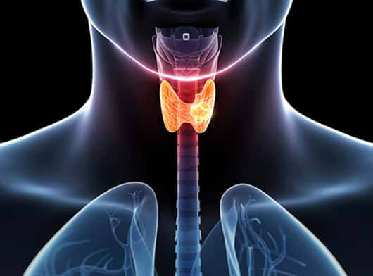
- Ultrasound – Ultrasound uses high-frequency sound waves to create pictures of body structures. For example, the ultrasound transducer is placed on the lower neck to create an image of the thyroid. This helps the doctor determine whether a thyroid nodule is likely to be benign or cancerous.
- Ultrasound-guided Biopsy – Fine Needle Aspiration Cytology (FNAC) – Ultrasound imaging typically uses the help of guiding a long thin needle through the skin and into the thyroid nodule to remove samples of suspicious thyroid tissue. This sample is then sent for further evaluation.
- Computed tomography (CT) scan – It is a diagnostic imaging procedure that uses x-rays to build cross-sectional images of the body. It also helps to identify the spread of cancer.
- Magnetic resonance imaging (MRI) – It is a diagnostic tool which uses the interaction of radio waves and magnetic field, which is processed in a high-speed computer system to produce detailed scan pictures of the tissue, organs, bones, ligament and cartilage. It may be useful in detecting tumours and their metastases. This diagnostic technique offers greater soft tissue contrast than a CT scan.
- PET CT– It is a diagnostic tool to assess spread to regional nodes or distant metastases to other parts of the body. It provides functional and morphological details by utilising radiation derived from Isotope labelled Glucose molecules to detect cellular glucose uptake in cancer. Post-surgery, it also helps to differentiate between scar tissue and tumour cells.
