


.png)


- Colonoscopy
- Imaging
- Laboratory
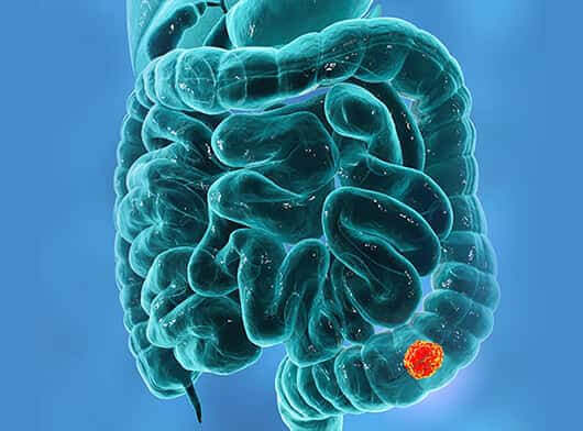
- Using an endoscope to examine the inside of the large intestine (colon) for abnormal changes. A long flexible tube is inserted into the rectum. A tiny video camera at the tip of the tube allows the doctor to view the inside of the colon
- Imaging -Tests such as CT and MRI are done to determine the stage of the cancer
- CT scan is a diagnostic imaging technique that uses x-rays to build cross-sectional images of the body.
- Magnetic Resonance Imaging (MRI) uses the interaction of radio waves and magnetic field which is processed in a high speed computer system to produce detailed scan pictures of the tissue, organs, bones, ligament and cartilage.
- CT/ Ultrasound guided Biopsy in this procedure CT scan/ Ultrasound is used to guide the needle into a suspected colorectal tumor in the safest possible manner. This is a minimally invasive procedure. The sample collected is sent for pathology evaluation.
- Blood tests - Tumor Markers – Carcino Embryonic Antigen (CEA) - CEA test is most often used to monitor colorectal cancer for people who are already receiving treatment. High levels of CEA may indicate that cancer has spread to other parts of the body. Other medical conditions can also cause CEA to increase.
- Pathology- The biopsy sample that is collected, is sent for further pathological evaluations such as Cytology, Immunohistochemistry (IHC) and Histopathological Evaluation (HPE).