


Direct answer
A CT scan (computed tomography, often called a "CT" or "cat scan") uses X-rays taken from many angles and a computer to build cross-sectional images of the body. CT is fast and detailed for bone, bleeding, chest, abdomen and blood vessels, but it uses ionising radiation and cannot reliably show every soft-tissue or early cartilage problem.
Key takeaways
- Strengths: CT is quick, widely available and strong for trauma, bleeding, lung disease, stones, abscesses and blood vessels.
- Limits: A normal CT does not mean disease is excluded; small, early or purely functional problems can be invisible on CT.
- Radiation: CT uses ionising radiation, so scans are done when the clinical question justifies them, at the lowest reasonable dose.
- Contrast matters: Iodinated contrast improves many studies but carries reaction and kidney-related considerations in some people.
- Not a substitute for MRI or ultrasound: Each answers a different question. Results vary depending on individual clinical circumstances.
At a glance
| Item | Detail |
|---|---|
| Test name | CT scan / computed tomography |
| Technology | X-rays (ionising radiation) plus computer reconstruction |
| Time in the scanner | Often under a minute of actual scanning; typically 5-15 minutes in the room, longer with contrast or multiple phases |
| Total visit time | Commonly 30-60 minutes including registration, cannula placement and observation after contrast |
| Contrast | May be intravenous, oral or rectal, depending on the study |
| Pain | The scan itself is painless; contrast injection may cause a warm flush or metallic taste |
| Report | Issued by a radiologist; must be interpreted alongside your symptoms and examination |
Also known as
- CT scan, CAT scan, "cat scan"
- Computed tomography, computerised tomography
- CECT (contrast-enhanced CT), NCCT (non-contrast CT)
- CT angiography (CTA), HRCT chest, CT KUB, CT brain (commonly written as "CT head plain")
- In Indian clinics you may hear "CT karwana hai", "CT plain" or "CT with dye"
What a CT scan is
A CT scanner is a ring-shaped machine. You lie on a motorised table that moves through the ring. Inside the ring, an X-ray tube and detectors rotate around you, collecting many projections. Software then reconstructs these into thin cross-sectional slices, which can be re-formatted into coronal, sagittal or three-dimensional views.
CT differs from a plain X-ray in that CT separates overlapping structures, so a liver, kidney, pancreas or brain haemorrhage can be seen individually rather than as overlapping shadows. Unlike MRI, CT does not use a magnetic field; unlike ultrasound, CT is not operator-dependent in image acquisition, but it does use radiation.
Why a CT scan is done
- Trauma: Head injury, suspected internal bleeding, solid-organ injury, spinal and complex fractures.
- Acute abdomen: Suspected appendicitis, bowel obstruction, perforation, diverticulitis, pancreatitis complications, abscess.
- Urinary stones: Non-contrast CT KUB for suspected ureteric colic.
- Chest disease: HRCT for interstitial lung disease and bronchiectasis; CT pulmonary angiography for suspected pulmonary embolism.
- Cancer care: Characterising a mass, staging, assessing treatment response, planning radiotherapy or surgery.
- Vascular questions: Aortic dissection or aneurysm, arterial blockage, venous thrombosis in selected sites.
- Stroke pathway: Non-contrast CT brain to look for haemorrhage before thrombolysis decisions, often with CT angiography and perfusion in stroke-ready centres.
- Guided procedures: CT-guided biopsy or drainage of collections.
Who should get this test
CT is a directed investigation, not a general health check. A CT scan is usually appropriate when:
- A doctor has a specific clinical question that CT can answer, such as suspected bleeding, stone, embolism, abscess, obstruction or tumour.
- Significant trauma has occurred, particularly head, chest, abdominal or spinal injury.
- An ultrasound, X-ray or blood test has raised a question that needs cross-sectional clarification.
- Cancer treatment requires staging or response assessment at planned intervals.
- A biopsy or drainage needs image guidance.
CT is usually not the right first step for undifferentiated fatigue, vague long-standing aches, routine screening in a person with no symptoms or risk factors, or as a "full body scan" package. Low-dose CT lung cancer screening is offered only to defined high-risk groups after a discussion of benefits and harms, and eligibility criteria differ between guidelines. Discuss with your doctor whether you fall into such a group.
What a CT scan cannot detect or exclude
CT is detailed, but it is not all-seeing. Being clear about the gaps prevents false reassurance.
- A normal CT does not exclude disease. It means no abnormality was identified on the images obtained, with the technique used, on that day.
- Early ischaemic stroke is often invisible on a non-contrast CT brain in the first hours. MRI with diffusion-weighted imaging is far more sensitive for early infarction.
- Coronary artery disease and angina: A routine chest CT does not assess the coronary arteries. Even CT coronary angiography shows anatomy, not whether a narrowing is limiting blood flow during exertion, that needs functional testing.
- Bone marrow, spinal cord, ligaments, cartilage, early bone oedema and many subtle brain lesions are better shown by MRI. Some fractures, including scaphoid, sacral insufficiency and some hip fractures, can be missed on CT and visible on MRI.
- Mucosal lesions of the gut: CT can miss small polyps, early gastric or colonic cancers and inflammatory changes limited to the lining. Endoscopy or colonoscopy remains the reference test for the mucosa.
- Small lung nodules, small liver and pancreatic lesions may be below the threshold of detection or may not be characterisable; follow-up or a different modality is often needed.
- Liver inflammation and fibrosis: CT can suggest fatty change or cirrhosis morphology but cannot grade inflammation or fibrosis. That requires elastography, blood-based scores or biopsy. Steatotic liver disease terminology has been updated internationally to MASLD (formerly NAFLD), MASH (formerly NASH) and MetALD where alcohol intake is also relevant.
- Function: CT shows structure. It does not tell you how well a kidney filters, how well a lung transfers oxygen or whether a nodule is metabolically active, nuclear medicine, PET-CT or physiological tests answer those questions.
- Benign versus malignant: CT often cannot settle this. Tissue diagnosis by biopsy is frequently required.
If your symptoms persist or worsen despite a normal CT report, go back to the doctor who requested the scan. A normal scan is not a reason to stop investigating an ongoing problem.
Related but different examinations
CT versus MRI
CT uses X-rays and is fast, making it suited to emergencies, bone detail, lung parenchyma, calcification and acute bleeding. MRI uses magnetic fields, takes longer, and gives superior contrast for brain tissue, spinal cord, marrow, ligaments, cartilage, pelvic organs and liver lesion characterisation. Metallic implants and devices require an MRI safety assessment rather than automatic exclusion.
CT versus ultrasound
Ultrasound involves no radiation, is inexpensive and is often the first test for gallbladder disease, many paediatric abdominal problems, thyroid and scrotal lesions, and anything in pregnancy. Ultrasound is limited by gas, body habitus and operator skill, and it cannot see much of the retroperitoneum or the mid-ureter reliably. USG abdomen and CT abdomen answer different questions and are frequently complementary.
CT versus plain X-ray
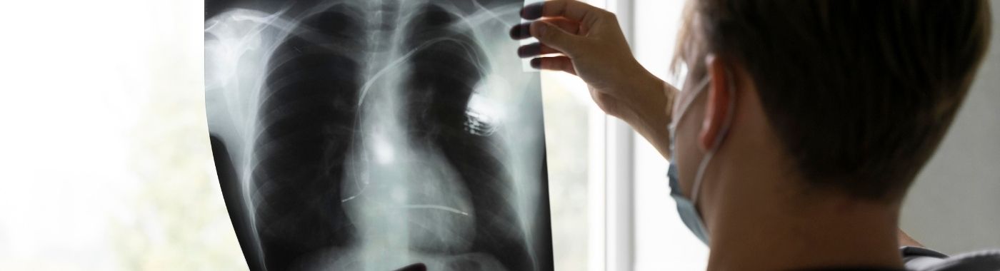
An X-ray is faster, cheaper and lower-dose, and remains adequate for many limb fractures, chest screening and device position checks. CT is used when an X-ray is normal or inconclusive but clinical concern persists, or when three-dimensional detail is needed.
Plain CT versus contrast-enhanced CT versus CT-guided intervention
These are not interchangeable. A plain (non-contrast) CT is used for suspected bleeding, head injury and urinary stones. A contrast-enhanced CT is needed to characterise most tumours, infections, vascular problems and solid-organ injury. CT angiography uses tightly timed contrast for arteries. A CT-guided biopsy or drainage is an interventional procedure with its own consent, bleeding and infection risks.
CT versus PET-CT
PET-CT adds a radiotracer to show metabolic activity, mainly in cancer staging and response assessment. PET-CT involves additional radiation and additional preparation, and it is not a better version of CT for most non-oncological questions.
How to prepare
Preparation is set by the individual radiology unit and depends on the body part, the scanner and whether contrast is planned. Fasting durations, oral contrast volumes, water intake instructions and arrival times vary between facilities. Your own unit's written instructions take precedence over anything you read here.
- Tell the unit in advance: any previous reaction to iodinated contrast, asthma, kidney disease or dialysis, diabetes and its medicines, thyroid disease, myeloma, pregnancy or breastfeeding, and all current medicines and supplements.
- Fasting: often requested for a few hours before a contrast-enhanced abdominal study; frequently not required for a plain CT head or limb.
- Kidney function: some units check serum creatinine or eGFR before intravenous contrast, particularly in older adults, people with diabetes, or known kidney disease.
- Oral contrast: for some abdominal studies you may be asked to drink a diluted contrast solution over a period before scanning.
- Clothing and metal: wear loose clothes; jewellery, hairpins, coins, belts and underwired garments over the scan area are usually removed.
- Previous imaging: bring earlier CT, MRI, ultrasound films and reports, comparison changes interpretation.
- Do not stop, start or change any prescribed medicine on your own. If a medicine needs to be adjusted around contrast, the prescriber or the radiology team will tell you.
If you are or might be pregnant
Tell the staff before entering the room. CT involves ionising radiation, so pregnancy usually prompts a review of whether ultrasound or MRI can answer the question. CT is still performed in pregnancy when the clinical need is compelling, such as suspected pulmonary embolism or major trauma, using dose-reduction techniques and after discussion of risks and benefits.
If you have diabetes on glucose-lowering therapy
Fasting instructions can affect blood sugar, particularly with insulin or sulfonylureas. Ask for an early appointment slot, carry your glucose monitor and a sugar source, and ask your prescriber how to handle your doses around the fasting period. Metformin is sometimes reviewed around iodinated contrast in people with reduced kidney function, that decision belongs to your treating doctor, not to you.
If you have chronic kidney disease or are on dialysis
Kidney function influences whether intravenous contrast is used, how much, and whether hydration protocols are applied. People on established dialysis are managed differently from those with declining but preserved function. Bring recent kidney reports.
If you have heart failure or a fluid restriction
Blanket instructions to "drink plenty of fluids" are unsafe for some people. If you have heart failure, advanced kidney disease or a prescribed fluid limit, ask the radiology team and your treating doctor for a hydration plan specific to you.
Children
Paediatric CT uses weight-adjusted, dose-reduced protocols, and alternatives such as ultrasound or MRI are considered first wherever they can answer the question. Very young children may need sedation to stay still; that requires separate assessment, fasting rules and monitoring.
Older adults
Mobility, hearing, cognition and kidney function all matter. A relative or attendant may be allowed in the control area. Transfers on and off the table should be assisted.
Claustrophobia and large body size
The CT ring is short and open at both ends, so CT is usually much better tolerated than MRI by people with claustrophobia. Table weight limits and gantry bore size vary between scanners; ring the unit beforehand if this may be relevant.
What happens during a CT scan
- Check-in and consent: your details, the request and your history are reviewed. For contrast studies, an intravenous cannula is placed, usually in the arm.
- Positioning: you lie on the table, usually on your back. Pillows or straps help you stay still.
- Scanning: the table moves through the ring. You may hear whirring or clicking. You may be asked to hold your breath for a few seconds for chest and abdominal scans.
- Contrast injection: if used, the injection often produces a warm flush, a metallic taste or a sensation of needing to pass urine. These pass within a minute. Tell the staff immediately about itching, rash, breathlessness, throat tightness or pain at the injection site.
- Staff contact: the radiographer works from an adjacent room with a glass window and an intercom, and can see and hear you throughout.
- Afterwards: most people leave and return to normal activity. After contrast, units often ask you to wait a short while for observation, and the cannula is removed before you go.
How long does the process take
The actual X-ray acquisition for most modern CT studies takes seconds to under a minute. Expect roughly 5-15 minutes on the table for a routine single-region study, and longer for multiphase studies, CT angiography, oral contrast preparation or CT-guided procedures. Including registration, cannulation and post-contrast observation, plan for about an hour in the department. Emergency scans are prioritised and can be completed much faster.
Understanding your CT report
A radiologist reviews the images and issues a structured report. Most reports have a technique section, a description of findings organ by organ, and an impression or conclusion. The impression is the part that answers the referring doctor's question.
- "No significant abnormality detected": nothing abnormal was identified within the limits of that technique. This is not the same as "you are free of disease".
- "Cannot be excluded" or "cannot be fully characterised": honest radiological language meaning the images do not settle the question. Further imaging, follow-up or biopsy may be advised.
- Incidental findings: small cysts, benign-looking nodules, gallstones, disc changes or fatty liver are often found unexpectedly and may need nothing more than noting. Many require no treatment.
- Descriptive terms: words like "hypodense", "enhancing", "ground-glass" or "stranding" describe appearances, not diagnoses.
- Follow-up recommendations: reports may suggest a repeat scan at an interval. Do not act or panic on the basis of a single line; ask the requesting doctor what it means for you.
There is no numeric "normal range" for a CT scan. Interpretation depends entirely on the region scanned, the clinical question and your history. Only the doctor who requested the scan, with your examination and other results in hand, can tell you what the report means for your care.
When CT results can be misleading
Findings that can be falsely suggested or exaggerated
- Movement and breathing artefact can blur images and mimic lesions or dissection flaps.
- Metal hardware, dental work and surgical clips create streak artefact that can look like an abnormality or hide a real one.
- Beam-hardening and partial volume effects near the skull base and bone can mimic haemorrhage or a lesion.
- Bowel gas, collapsed bowel loops or inadequate oral contrast can imitate a mass or thickening.
- Inflammation after recent surgery, infection or radiotherapy can look like residual or recurrent tumour.
- Incidental nodules and cysts are common and frequently benign, yet can trigger anxiety and further testing.
Findings that can be falsely reduced or missed
- Wrong timing or absent contrast: a plain scan can miss vascular lesions, abscesses and many tumours that only become visible with contrast at the correct phase.
- Very early disease: early stroke, early osteomyelitis and early marrow infiltration can appear normal on CT.
- Small lesions below the slice-resolution threshold, or lesions with the same density as the surrounding organ, can be invisible.
- Anaemia or altered haematocrit changes blood density, so acute haemorrhage can look less obvious than expected.
- Region not covered: a CT chest does not evaluate the lower abdomen. If the symptomatic area was outside the scan field, it was not assessed.
- Dose-reduced protocols, appropriately used in children and pregnancy, are noisier and may reduce sensitivity for subtle findings.
Risks and safety
CT is widely used and generally well tolerated, but the risks differ by the type of study. Results and risks vary depending on individual clinical circumstances.
Plain (non-contrast) CT
The main consideration is radiation. Dose varies by body region, scanner, protocol and body size. Individual risk from a single medically indicated CT is small, and for an acute problem the information usually outweighs it. Risk accumulates with repeated scanning, which is why unnecessary or duplicate scans are avoided and prior imaging is used for comparison. Children are more radiosensitive and have longer remaining lifespan, so paediatric protocols are dose-optimised.
Contrast-enhanced CT with intravenous iodinated contrast
- Common, transient effects: warmth, flushing, metallic taste, mild nausea.
- Hypersensitivity reactions: usually mild, such as itching or urticaria. Severe reactions including bronchospasm and anaphylaxis are uncommon but possible, which is why contrast is given in units equipped to manage them.
- Kidney effects: post-contrast acute kidney injury is a recognised concern, mainly in people with significantly reduced kidney function, dehydration or multiple risk factors. Modern evidence suggests the risk in people with normal kidney function is lower than once believed, but assessment is still individualised.
- Extravasation: contrast leaking into arm tissues causes swelling and pain; report this immediately.
- Thyroid: iodinated contrast can affect thyroid function tests and thyroid treatment planning, so mention thyroid disease or planned radioiodine therapy.
- Breastfeeding: only very small amounts pass into milk; most guidance supports continuing breastfeeding, but confirm with your unit.
Oral and rectal contrast
Usually causes only bloating, altered taste or loose stools. Aspiration risk is relevant in people with swallowing difficulty or reduced consciousness; tell staff if this applies.
CT-guided biopsy and drainage
These are invasive procedures with additional risks including bleeding, infection, pain, pneumothorax for chest procedures, and injury to adjacent structures. Separate consent, blood-thinner review and post-procedure observation apply.
Sedation or anaesthesia
Where sedation is used, mainly in young children or people unable to lie still, standard sedation risks and monitoring requirements apply.
Red flags
Emergency: seek care now
- During or after contrast: breathlessness, wheeze, throat or tongue swelling, widespread rash, faintness or collapse.
- Sudden severe chest pain, tearing back pain, sudden one-sided weakness, facial droop, slurred speech or sudden loss of vision.
- Sudden severe headache described as the worst ever, with vomiting or reduced consciousness.
- Coughing blood with breathlessness, or sudden severe abdominal pain with a rigid abdomen.
- Following a CT-guided chest procedure: increasing breathlessness or sharp chest pain.
Same-day medical review
- Marked swelling, blistering or increasing pain at the contrast injection site.
- Rash, itching or facial puffiness developing in the hours after contrast.
- Passing much less urine than usual after contrast, particularly with known kidney disease.
- New fever, persistent vomiting or worsening pain after a CT-guided biopsy or drainage.
- A report you have been told contains an urgent finding that needs immediate action.
Routine appointment
- Discussing a report that mentions an incidental nodule, cyst or recommended follow-up interval.
- Ongoing or worsening symptoms despite a normal CT, this needs review, not reassurance.
- Questions about cumulative radiation from repeated scans, or about whether ultrasound or MRI could be used next time.
- Mild transient nausea or a metallic taste that settled on its own.
Special situations
Pregnancy
Abdominal and pelvic CT is avoided where a reasonable alternative exists. Where CT is genuinely needed, foetal dose is minimised and the decision is shared. In India, CT is not a prenatal sex-determination tool; any imaging in pregnancy is performed strictly for clinical indications.
Children
Ultrasound and MRI are preferred whenever they can answer the question. When CT is required, for example in significant head injury or major trauma, paediatric dose-reduction protocols are used, and scans are limited to the necessary region.
Older adults
Kidney function declines with age even when creatinine looks acceptable, so contrast decisions are individualised. Comorbidity and frailty influence whether an abnormal finding will change management at all, worth discussing before agreeing to further scans.
Diabetes on glucose-lowering therapy
The fasting period, not the scan, is the main hazard. Plan dose timing with your prescriber and carry treatment for low blood sugar.
Chronic kidney disease
Contrast may still be used when the diagnostic benefit is high, with hydration and dose adjustment. Alternatives such as ultrasound, non-contrast CT or MRI are considered.
Heart failure
Contrast volume and any hydration protocol must account for fluid tolerance. Tell the radiology team about your heart failure and any fluid restriction.
Implants, devices and pacemakers
Cardiac devices, stents and orthopaedic metal do not prevent a CT scan. They may create artefact. It is MRI, not CT, that requires a formal magnetic safety assessment.
CT scanning in the Indian context
- Terminology: requests are often written as "CT plain", "CECT", "CT KUB", "HRCT chest" or "CT brain". Ask which one you are booked for, and whether contrast is planned, because the preparation differs.
- Radiation regulation: CT installations in India operate under Atomic Energy Regulatory Board (AERB) requirements for registration, quality assurance and radiation protection. Staff wear monitoring badges and shielding is built into the room design.
- PC-PNDT Act, 1994: applies to prenatal diagnostic procedures and prohibits any use of imaging for foetal sex determination. Imaging in pregnancy is done only on clinical indication.
- Advertising law: the Drugs and Magic Remedies (Objectionable Advertisements) Act, 1954 and NMC professional conduct rules restrict exaggerated diagnostic claims. Be sceptical of "full body CT health package" marketing, whole-body screening CT in healthy people is not supported as a routine wellness measure and generates incidental findings that lead to further testing.
- Data protection: images, reports and any online form you fill in are personal health data governed by the Digital Personal Data Protection Act, 2023. Ask how your images are stored and shared.
- Clinical patterns that matter locally: tuberculosis is a common differential on chest and abdominal CT in India and can mimic malignancy; a CT appearance suggestive of TB usually still needs microbiological or tissue confirmation. Anti-tubercular therapy hepatotoxicity, hepatitis E, and liver injury from herbal, AYUSH or gym supplements are frequent reasons for abnormal liver findings. South Asians develop metabolic and fatty liver disease at lower body mass index, so a fatty liver seen incidentally on CT is common even in people who do not consider themselves overweight. Iron-deficiency anaemia is widespread and can alter the density of blood on CT.
- Bring your files: prior films and reports are often held by patients rather than centrally shared. Carrying them prevents repeat scanning and unnecessary radiation.
Cost and insurance in India
CT prices in India vary widely between cities, between government and private facilities, and between scanner generations. Costs are indicative only and change frequently; confirm the current charge with the imaging centre before booking, and ask for a written estimate that states whether contrast is included.
Factors that affect the price:
- Region and number of regions scanned: head, chest, abdomen, spine, or a combined study.
- Contrast: contrast-enhanced and angiographic studies cost more than plain scans because of the agent, the cannula, the injector and observation time.
- Number of phases: triple-phase liver or multiphase renal studies take more time and radiation planning.
- Scanner technology: dual-energy, high slice count and cardiac-capable scanners generally cost more.
- Special reconstructions: 3D, CT angiography, dental or radiotherapy planning reconstructions.
- Urgency and timing: emergency, night and holiday scans may be charged differently.
- Interventional CT: biopsy or drainage includes procedure, consumables, pathology and observation charges.
- Sedation or anaesthesia support: mainly for children.
Insurance: CT is generally reimbursable when clinically indicated during an admission or an approved day-care pathway. Outpatient diagnostic scans are covered only by some policies and riders. Government schemes such as Ayushman Bharat PM-JAY and various state schemes cover imaging under defined packages at empanelled facilities. Screening or self-requested "package" scans are usually not covered. Check your policy wording, sub-limits and pre-authorisation requirements with your insurer or the hospital's insurance desk before the scan.
Myths and facts
| Myth | Fact |
|---|---|
| A CT scan checks the whole body for any disease. | A CT scan covers only the region requested, and detects only what that technique can show. Many conditions need blood tests, endoscopy, MRI or functional testing instead. |
| A normal CT means nothing is wrong. | A normal CT means no abnormality was identified with that technique. Persisting symptoms still need review. |
| CT and MRI are the same thing, MRI is just newer. | They use different physics and answer different questions. CT is better for bone, lung and acute bleeding; MRI is better for cord, marrow, ligaments and brain tissue detail. |
| Contrast dye damages everyone's kidneys. | Contrast-related kidney injury is mainly a concern in people with significantly reduced kidney function or several risk factors. Most people with normal kidneys tolerate it. |
| One CT scan will give me cancer. | The individual risk from a single medically indicated CT is small. The concern is unnecessary and repeated scanning, which is why indications are reviewed. |
| Radiation stays in the body after a CT scan. | Diagnostic X-rays do not remain in the body. You are not radioactive after a CT scan and pose no risk to others. This differs from nuclear medicine and PET, which use injected radiotracers. |
| Everyone must fast before a CT scan. | Fasting is usually only needed for certain contrast studies. Many plain scans need no fasting. Follow your unit's instructions. |
| A "cat scan" involves a machine you are fully enclosed in. | The CT ring is short and open at both ends. It is usually far less confining than an MRI tunnel. |
Frequently asked questions
Is a CT scan painful?
The scan itself is painless. If contrast is injected, the cannula insertion stings briefly and the contrast can cause a warm flush, a metallic taste or a false urge to pass urine. These sensations settle within a minute. Tell the radiographer at once if you develop pain at the injection site.
How much radiation does a CT scan involve?
Dose depends on the body region, protocol, scanner and body size, so a single figure is misleading. A CT delivers more radiation than a plain X-ray and none from ultrasound or MRI. For a genuine clinical question, the diagnostic benefit usually outweighs the small individual risk.
Do I need to drink a lot of water after contrast?
Many units advise extra fluids after iodinated contrast to support kidney handling. That advice is not safe for everyone. If you have heart failure, advanced kidney disease or a prescribed fluid restriction, ask the radiology team and your treating doctor for a plan suited to you.
Can I have a CT scan while breastfeeding?
Yes in most cases. Only a very small amount of iodinated contrast passes into breast milk, and current guidance generally supports continuing breastfeeding without interruption. Confirm with your imaging unit, as local policies differ and some still advise a short pause.
Why did my doctor order a CT when I already had an ultrasound?
Ultrasound and CT answer different questions. Ultrasound is excellent for gallbladder, kidneys and superficial structures but is limited by gas, depth and body habitus. CT gives a








 Best Hospital Near me Chennai


