


Direct answer
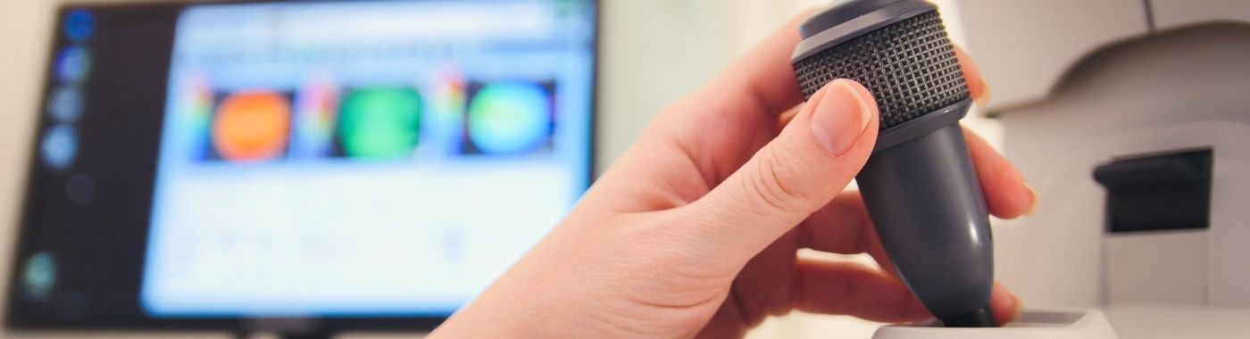
Corneal topography is a quick, non-contact scan that maps the curvature and shape of the cornea's front surface and presents it as a colour-coded map. Eye surgeons use corneal topography to screen for keratoconus, plan LASIK and other refractive surgery, fit contact lenses on irregular corneas, and follow corneal shape after surgery or injury.
Key takeaways
- Corneal topography maps corneal shape, not vision, nerve function or retinal health.
- Standard topography reads the front surface only; it does not measure corneal thickness or the back surface, that needs tomography (Scheimpflug or OCT-based).
- A normal topography map does not exclude early keratoconus, dry eye disease, glaucoma or retinal disease.
- Contact lens wear, dry eyes and poor fixation can distort the map and mimic or mask disease.
- Maps must be read alongside symptoms, refraction, corneal thickness and family history by the treating ophthalmologist.
At a glance
| Feature | Detail |
|---|---|
| What it examines | Curvature and elevation of the corneal surface |
| Type | Non-contact optical imaging; no needles, no dye, no radiation |
| Anaesthesia | None for standard topography |
| Time in the room | Usually a few minutes per eye; about 10-20 minutes including positioning, repeat captures and checks |
| Discomfort | Minimal; some people find the bright rings or flash uncomfortable briefly |
| Main uses | Keratoconus screening and monitoring, refractive surgery workup, complex contact lens fitting, post-surgical and post-transplant follow-up, astigmatism assessment before cataract surgery |
| Report availability | Maps are generated immediately; clinical interpretation happens at your consultation |
Also known as
- Videokeratography / videokeratoscopy
- Placido disc topography (the ring-pattern type)
- Corneal mapping or "eye mapping test"
- Topo or "topography test", the usual shorthand in Indian eye clinics
- Pentacam / Orbscan / Sirius / Galilei scan: patients often call the test by the machine's brand name. Several of these are actually corneal tomography devices, which do more than topography.
What corneal topography is
The cornea is the clear dome at the front of the eye and supplies most of the eye's focusing power. Small irregularities in its shape have a large effect on vision. Corneal topography measures that shape without touching the eye.
Two broad technologies are in common use:
- Reflection-based (Placido disc) topography: concentric lighted rings are projected onto the cornea and the reflected pattern is analysed. This measures the front surface only.
- Elevation-based tomography (rotating Scheimpflug camera, scanning slit, or anterior segment OCT): images cross-sections of the cornea, so it can also map the back (posterior) surface and produce a corneal thickness (pachymetry) map. Many Indian refractive clinics use these devices and still call the printout "topography".
The output is a colour-coded map. By convention, warmer colours (red, orange) mark steeper areas and cooler colours (blue, green) mark flatter areas. The colour scale is chosen by the operator, so the same eye can look dramatically different on two printouts. Colour alone is not a diagnosis.
Why corneal topography is done
- Refractive surgery screening: to look for shape patterns that suggest keratoconus or other ectasia, which would make LASIK or SMILE unsafe.
- Keratoconus detection and monitoring: to identify irregular steepening and to compare serial scans for progression.
- Astigmatism assessment: to characterise the amount, axis and regularity of astigmatism, including before cataract surgery when a toric lens is being considered.
- Contact lens fitting: especially rigid gas-permeable, scleral and specialty lenses on irregular or post-surgical corneas.
- After corneal procedures: following cross-linking, refractive surgery, corneal transplant, ring segment implantation or corneal injury.
- Unexplained blurred vision: when spectacle correction does not give clear vision, or when the refraction keeps changing.
Who should get this test
- Anyone being assessed for LASIK, PRK, SMILE or a phakic intraocular lens.
- People with rapidly changing spectacle power, worsening astigmatism, or glasses that no longer sharpen vision.
- Children, teenagers and young adults with a family history of keratoconus, or with persistent eye rubbing and allergic eye disease, a common and important combination in India.
- People already diagnosed with keratoconus, for periodic monitoring at the interval their ophthalmologist advises.
- People needing rigid or scleral contact lenses.
- Patients planned for cataract surgery where astigmatism correction is being considered.
- Those with corneal scarring, past corneal infection, corneal transplant, or trauma affecting the cornea.
- People with keratoconus-associated conditions such as Down syndrome, or connective tissue disorders, when advised by the treating ophthalmologist.
Corneal topography is not a general eye check-up and is not needed by everyone. It is ordered for a specific question. Results vary depending on individual clinical circumstances.
What corneal topography cannot detect or exclude
Corneal topography answers one question: what shape is the corneal surface? It does not assess the rest of the eye.
- It does not measure vision or spectacle power. Refraction and visual acuity testing are separate.
- Standard Placido topography does not measure corneal thickness and does not image the posterior corneal surface. Early ectasia can begin posteriorly, so front-surface-only maps can look acceptable while tomography shows abnormality.
- A normal map does not exclude early or subclinical keratoconus. Some eyes that later progress look normal on the first scan. Serial scans over time are often what settle the question.
- It does not diagnose dry eye disease, though a poor tear film degrades the map.
- It does not assess the retina, macula, optic nerve or intraocular pressure. Glaucoma, diabetic retinopathy, macular disease and optic neuropathy cannot be found or ruled out on topography.
- It does not diagnose infection or inflammation. Corneal ulcers, keratitis and uveitis need slit-lamp examination and, when relevant, microbiological testing.
- It does not count endothelial cells (that is specular microscopy) or measure axial length for lens power calculation (that is biometry).
- It does not predict, on its own, whether refractive surgery will succeed. Decisions combine topography or tomography with pachymetry, refraction stability, pupil size, dry eye status and ocular surface health.
A finding that is "not identified" on a topography map means it was not seen on this scan, in these conditions, on this device. It does not mean the condition is absent. If your vision continues to change or blur despite a normal map, go back to your ophthalmologist rather than assuming the eye has been cleared.
Related but different examinations
| Test | Question it answers | Key difference from corneal topography |
|---|---|---|
| Corneal tomography (Scheimpflug, scanning slit, anterior segment OCT) | What is the shape of both corneal surfaces and how thick is the cornea across its area? | Adds posterior surface and full pachymetry mapping; generally preferred for ectasia screening before refractive surgery |
| Keratometry (manual or auto) | What are the two principal curvatures in the central cornea? | A few data points only; cannot show irregular or off-centre steepening |
| Pachymetry (ultrasonic or optical) | How thick is the cornea? | Thickness only, no shape map; ultrasonic pachymetry does touch the eye and needs anaesthetic drops |
| Ocular wavefront aberrometry | How does the whole optical system, including the lens, distort light? | Measures the optics of the entire eye, not corneal shape alone |
| Specular (endothelial) microscopy | Are the endothelial cells on the inner corneal surface healthy and adequate in number? | Cell layer health, not curvature |
| Optical biometry | What intraocular lens power is needed for cataract surgery? | Measures axial length and anterior chamber depth; uses keratometry values but is not a shape map |
| Retinal OCT / fundus imaging | Is the macula, retina or optic nerve affected? | Back of the eye; entirely different structures from the cornea |
These tests are complementary, not ranked. Which one comes first depends on the clinical problem. For a young person with worsening astigmatism and suspected keratoconus, tomography with a pachymetry map is usually the more informative starting point. For an established keratoconus patient being fitted with a scleral lens, surface topography may be what the fitter needs. For blurred vision with a clear cornea and normal shape, the priority shifts to retinal and optic nerve assessment instead.
How to prepare
Preparation protocols differ between eye units and devices. Your clinic's written instructions take precedence over anything you read here.
- Contact lenses: lenses temporarily reshape the cornea, so a wash-out period is usually advised before topography, especially before refractive surgery planning. The period depends on lens type, lens material, how long you have worn lenses and why the scan is being done; it can range from a few days for occasional soft lens wear to several weeks for rigid gas-permeable or long-term wear. Ask your clinic for a specific instruction, and do not stop a medically prescribed therapeutic lens without speaking to the prescriber.
- Eye drops: tell the clinic about all drops you use, including lubricants, glaucoma drops, steroids and antibiotics. Do not stop any prescribed drop on your own, check with the prescriber.
- Eye rubbing: avoid rubbing your eyes on the day of the test. Vigorous rubbing can distort the surface briefly and also contributes to keratoconus progression over time.
- Eye make-up and creams: many units ask you to attend without eye make-up, kajal or oily creams, as debris on the lids and tear film degrades image quality.
- Dry eyes: if your eyes feel gritty or burn, mention this before the scan. Some units treat the surface first and rescan, because a poor tear film is a leading cause of unreliable maps.
- Bring your records: old spectacle prescriptions, previous topography printouts and details of any earlier eye surgery help enormously with comparison.
If you have had recent eye surgery or injury
Corneal shape continues to change for weeks to months after refractive surgery, cataract surgery, cross-linking, corneal transplant or a significant abrasion. Scans taken too early may be unrepresentative. Your surgeon decides the timing.
Children and adolescents
Topography needs a few seconds of steady fixation and a chin-and-forehead rest. Many school-age children manage well; younger or anxious children may need repeated attempts, a handheld device where available, or examination under sedation in selected cases. Children with allergic eye disease who rub their eyes are an important group to screen, and repeat scans are often needed as results in children are more likely to be unreliable on the first attempt.
Older adults and people with reduced mobility
Positioning at the chin rest can be difficult with neck or back problems, tremor or a wheelchair. Tell the technician in advance; alternative seating or a device with more flexible positioning may be available.
Pregnancy
Corneal topography uses light only, no radiation and no contrast, so there is no pregnancy-specific hazard from the scan itself. However, corneal shape and refraction can shift during pregnancy and breastfeeding. For this reason refractive surgery planning scans are usually deferred until the measurements are stable. Discuss timing with your ophthalmologist.
Severe dry eye, allergy or ocular surface disease
These conditions reduce map reliability in a predictable way. Treating the surface first and then rescanning gives a more trustworthy result than acting on a poor-quality initial map.
What happens during corneal topography
- Check-in: your details, spectacle power and lens-wear history are recorded.
- Positioning: you sit facing the device with your chin on a rest and forehead against a band. The technician aligns the instrument to your eye.
- Capture: you are asked to blink normally, then keep your eye open wide and stare at a small central target. Lighted rings or a rotating camera pass across the eye. Each capture takes a second or two. Nothing touches the eye.
- Repeats: several captures per eye are usual. The technician checks quality scores and may repeat scans if blinking, tears or lash shadows interfere. Being asked to repeat the scan is normal and does not mean something is wrong.
- Both eyes: each eye is scanned separately, and comparing the two eyes is an important part of interpretation.
How long the process takes
The imaging itself is very short, seconds per capture. Allow roughly 10-20 minutes for the whole appointment including positioning, repeat captures and quality checks. If the scan is part of a refractive surgery workup with refraction, dilated examination, pachymetry and a surgeon consultation, expect to be at the clinic for a few hours. Total time varies between units and depends on how many additional tests are combined.
Understanding your report
A topography report typically includes a colour map of curvature, keratometric readings for the steepest and flattest meridians with their axes, indices summarising regularity and asymmetry, and a quality or reliability score. Tomography reports add posterior elevation maps and a thickness map.
- Regular, symmetrical pattern: curvature changes smoothly and the two eyes look broadly similar. Regular astigmatism often appears as a symmetrical bow-tie.
- Asymmetric or localised steepening: an inferior or off-centre steep zone, a skewed bow-tie, or a large difference between the two eyes may raise suspicion of keratoconus or another ectasia. Suspicion is not diagnosis, it prompts thickness assessment, examination and repeat scans.
- Flattening after surgery: expected after myopic LASIK or PRK; the central cornea is deliberately flatter.
- Irregular islands or distorted rings: may reflect scarring, a healed ulcer, previous trauma, pterygium encroaching on the cornea, or simply a poor tear film.
- Indices and "probability" scores: device software may print a keratoconus index or risk percentage. These are statistical tools derived from specific populations and devices. They are screening aids, not verdicts, and they generate both false alarms and missed cases.
Grading and classification on topography is partly interpretive. Colour scales, device model, software version and normative database all affect appearance and indices, so the same eye can be labelled differently in two clinics. Serial scans should ideally be done on the same machine for comparison. No topography report is a diagnosis on its own, it must be interpreted in context by the ophthalmologist who requested it.
When results can be misleading
Factors that can make the cornea look more abnormal than it is
- Dry eye or a broken-up tear film: the commonest cause of spuriously irregular maps.
- Recent contact lens wear: especially rigid lenses, which can leave corneal warpage that mimics ectasia and resolves over weeks.
- Eye rubbing shortly before the scan.
- Poor fixation, blinking mid-capture, or misalignment: produces artefactual steep or flat zones.
- Lash or lid shadows and a narrow palpebral aperture: data drop-out in the periphery may be extrapolated by the software.
- Pterygium, corneal scar, dystrophy or Salzmann nodules: local surface change interpreted as ectasia.
- Over-tight colour scaling: a normal cornea can look alarming when the scale is narrow.
Factors that can make the cornea look more normal than it is
- Front-surface-only devices in early disease: posterior changes and focal thinning are missed.
- Prior myopic refractive surgery: flattening can mask underlying steepening, making post-LASIK ectasia harder to spot on curvature maps alone.
- Corneal oedema or scarring that smooths the surface.
- Averaging of multiple captures, which can dilute a small focal abnormality.
- A single scan in a young patient: progression is defined by change over time, so one normal scan cannot exclude evolving keratoconus.
When the map and the clinical picture disagree, the clinical picture and repeat testing usually win.
Risks and safety
Standard non-contact topography and tomography
These scans use visible light or near-infrared light, involve no contact with the eye, no dye and no ionising radiation. Reported problems are limited to brief dazzle from the illumination, transient after-images, and mild discomfort from holding the eye open or leaning on the chin rest. There is no meaningful infection risk when the forehead and chin rests are cleaned between patients. People with photosensitivity, migraine or epilepsy triggered by flashing light should mention this beforehand.
Contact-based measurements sometimes done in the same sitting
Not every test in a corneal workup is non-contact. Ultrasonic pachymetry and applanation tonometry touch the cornea after anaesthetic drops, and carry a small risk of corneal abrasion, drop stinging and, rarely, allergic reaction to the anaesthetic. Safety statements for non-contact topography should not be assumed to cover these. Ask which of your tests involve contact.
Dilating drops
If dilating drops are used for other parts of the examination, expect blurred near vision and light sensitivity for several hours, and arrange not to drive. Dilating drops are used with caution in people with very narrow anterior chamber angles.
Red flags: when to seek help rather than wait for a scan
Corneal topography is an elective imaging test, not an emergency service. If any of the following apply, the priority is examination, not imaging.
Emergency: seek eye care now
- Sudden loss of vision, or a curtain or shadow across the vision.
- Severe eye pain with redness, marked light sensitivity, vomiting or a fixed dilated pupil.
- Penetrating injury, chemical splash, or anything embedded in the eye.
- A white or grey spot on the cornea with pain and discharge, particularly in a contact lens wearer, possible corneal ulcer.
- Sudden double vision, drooping eyelid or new squint with headache.
Same day or within 24-48 hours
- New floaters with flashes of light.
- Rapidly worsening blurring over days, or sudden onset of distorted vision in one eye.
- A painful red eye that is not settling, or increasing discharge and lid swelling.
- Any pain, sudden haze or vision drop after recent eye surgery, cross-linking or a corneal transplant, this could signal graft rejection or infection.
Routine appointment
- Spectacle power that keeps changing, or glasses that no longer give clear vision.
- Gradually increasing astigmatism, glare, halos or ghosting of images.
- Persistent eye rubbing and itching, especially in a child with allergy.
- Family history of keratoconus and no previous screening.
- Considering LASIK, SMILE or PRK.
- Follow-up monitoring of known keratoconus at the interval your ophthalmologist advises.
Special situations
Keratoconus suspects and young patients
In teenagers and people in their twenties, keratoconus can progress quickly. A single scan often cannot settle the diagnosis; repeat scans at intervals chosen by the ophthalmologist, ideally on the same device, are what demonstrate stability or progression. Reducing eye rubbing and treating allergic eye disease are central parts of management.
Before and after refractive surgery
Screening scans identify eyes at higher risk of post-operative ectasia, and some eyes are advised against corneal laser surgery on this basis. Being declined is a safety decision, not a failure of the test. After surgery, scans track the new shape and help explain residual blurring, glare or regression. Outcomes vary depending on individual clinical circumstances.
Cataract surgery
Topography or tomography helps decide whether astigmatism is regular enough for a toric intraocular lens. Irregular corneas, dry eye and previous refractive surgery all change the calculation and the accuracy of predicted results.
Contact lens wearers
If you wear lenses, expect a wash-out period before diagnostic scans. Never stop a therapeutic or prosthetic lens prescribed for a medical reason without speaking to the prescribing doctor.
Corneal transplant recipients
Maps after keratoplasty are typically irregular, with steep and flat sectors related to sutures and wound healing. Suture adjustment, later removal and lens fitting are guided partly by these maps. Comparison with previous scans matters more than a single printout.
People with systemic conditions
Diabetes affects the corneal surface and tear film and can reduce map reliability. Connective tissue disorders and Down syndrome are associated with a higher likelihood of keratoconus, so screening may be advised earlier and repeated.
Corneal topography in the Indian context
- Keratoconus is clinically important in India. Indian tertiary eye centres report keratoconus commonly in adolescents and young adults, frequently alongside allergic eye disease and habitual eye rubbing, and often at a younger age and with more advanced disease at presentation than in many Western series. Early topographic screening in a child with persistent itching and rubbing is therefore worthwhile.
- Allergic conjunctivitis and vernal keratoconjunctivitis are widespread in Indian children. Both worsen the tear film and drive eye rubbing, so they reduce scan reliability and raise keratoconus risk at the same time.
- Dry eye and environmental factors: urban air pollution, dust, long screen hours and air-conditioned workplaces all destabilise the tear film. Lubrication and surface treatment before scanning often improve map quality.
- Kajal, surma and kohl deposit on the lid margin and tear film, and can degrade image quality. Attend without eye cosmetics if your clinic asks.
- Device naming: patients frequently ask for a "Pentacam" or "Orbscan" by name. Ask your clinic which device is being used and, for serial monitoring, try to stay with the same device and centre so the comparison is valid.
- Brand and clinic variation: availability of Scheimpflug tomography, epithelial thickness mapping and biomechanical testing differs widely between metros, tier-2 cities and smaller towns. Where advanced ectasia screening is not available locally, referral to a corneal service is reasonable before any laser refractive procedure.
- Data privacy: corneal scans and the personal details attached to them are health data. Under the Digital Personal Data Protection Act 2023, any digital form, appointment tool or report portal that collects them requires clear notice and your consent for the stated purpose.
- Advertising standards: claims about laser vision correction outcomes are constrained by the Drugs and Magic Remedies (Objectionable Advertisements) Act 1954 and by NMC professional conduct rules. Be cautious about any package promising a guaranteed visual outcome.
Cost and insurance in India
Charges for corneal topography in India are usually modest compared with most imaging tests, and typically fall in the low hundreds to low thousands of rupees per session. Advanced tomography, dual-device protocols and epithelial or biomechanical add-ons cost more. Figures move frequently and differ by city and centre, so ask for a written estimate before booking.
What drives the cost:
- Test type: basic Placido topography versus Scheimpflug or OCT-based tomography; whether pachymetry and elevation maps are included.
- One eye or both eyes, and whether serial monitoring scans are billed separately.
- Bundling: refractive surgery and cataract workups often package topography with refraction, biometry, tonometry and consultation, which changes the apparent price.
- Type of facility: standalone optical or optometry clinic, single-specialty eye hospital, or multi-specialty tertiary centre.
- City tier and competition in the local market.
- Who interprets the report and whether a corneal specialist consultation is included.
- Repeat scans needed after a contact lens wash-out or after treating dry eye.
Insurance: diagnostic tests done purely as outpatient investigations are often not covered unless your policy includes OPD benefits. Topography done as part of a covered admission, for example a cataract surgery workup, is more commonly reimbursed. Refractive surgery for spectacle removal is usually treated as elective or cosmetic and excluded, though some policies cover it above a defined refractive power or after a waiting period. Corneal cross-linking and keratoplasty for disease are more often covered than elective laser correction. Check your specific policy wording and pre-authorisation requirements with the insurer or the hospital's insurance desk before the procedure.
Myths and facts
| Myth | Fact |
|---|---|
| Corneal topography is a complete eye check-up. | Corneal topography examines corneal shape only. It says nothing about the retina, optic nerve or eye pressure. |
| A normal topography rules out keratoconus. | Early keratoconus can be missed, particularly on front-surface-only devices. Repeat scans and thickness mapping are often required. |
| Red on the map means disease. | Red simply marks steeper areas on the chosen colour scale. Normal steep corneas appear red; the scale itself can be adjusted. |
| The test is painful or involves an injection. | Standard corneal topography is non-contact, with no needles, dye or radiation. Some people find the lights briefly uncomfortable. |
| Topography improves vision. | Corneal topography is diagnostic only. Any improvement comes from the lenses, cross-linking or surgery it helps plan. |
| Topography and tomography are the same thing. | They overlap but differ. Tomography also images the posterior cornea and maps thickness, which matters for ectasia screening. |
| If topography is normal, LASIK is certainly safe. | Suitability depends on corneal thickness, refraction stability, ocular surface health, pupil size and other factors, not the shape map alone. |
| A high keratoconus index on the printout means you have keratoconus. | Software indices are screening aids with false positives and false negatives. Diagnosis requires clinical correlation. |
Frequently asked questions
Does corneal topography hurt, and does anything touch my eye?
Standard corneal topography and tomography are non-contact. Lights or a rotating camera are directed at the eye and nothing touches the surface. Most people report only brief dazzle or mild strain from holding the eye open. Other tests in the same visit, such as ultrasonic pachymetry or applanation tonometry, do touch the cornea after anaesthetic drops.
How long does the whole appointment take?
Each capture lasts a second or two, and a few captures per eye are usual. Plan for about 10-20 minutes for topography alone. If it forms part of a refractive surgery or cataract workup with refraction, dilation and a surgeon consultation, allow several hours at the clinic. Timings vary between units.
How long must I stop contact lenses before the scan?
Contact lenses temporarily reshape the cornea, so a wash-out period is usually needed. The length depends on lens type, material, duration of wear and the purpose of the scan, ranging from days to several weeks. Your eye unit will give you a specific instruction. Do not stop a therapeutic lens without asking the prescribing doctor.
Can corneal topography diagnose keratoconus on its own?
Corneal topography is a central tool for suspecting and monitoring keratoconus, but diagnosis combines the map with corneal thickness, slit-lamp examination, refraction, symptoms and family history. Posterior surface and pachymetry data from tomography often add decisive information. A single normal scan does not exclude early disease.
Why has my clinic asked me to repeat the scan?
Repeats are routine. Blinking, a dry or unstable tear film, lash shadows or slight misalignment lower the quality score, and technicians rescan to obtain reliable data. A repeat request usually reflects image quality rather than an abnormal finding. Repeat scanning after treating dry eye commonly produces a cleaner map.
What is the difference between topography and a Pentacam or Orbscan scan?
Pentacam and Orbscan are brand names for corneal tomography devices. Tomography images cross-sections of the cornea, so it maps the posterior surface and corneal thickness in addition to front curvature. Classic Placido topography measures the front surface only. For ectasia screening before laser surgery, tomographic data are generally preferred.
Is corneal topography safe in pregnancy and for children?
The scan uses light only, with no radiation or contrast, so it poses no pregnancy-specific hazard. However, corneal measurements can shift during pregnancy and breastfeeding, so refractive surgery planning is usually deferred. Children often cooperate well, though fix









 Best Hospital Near me Chennai


