


Директан одговор
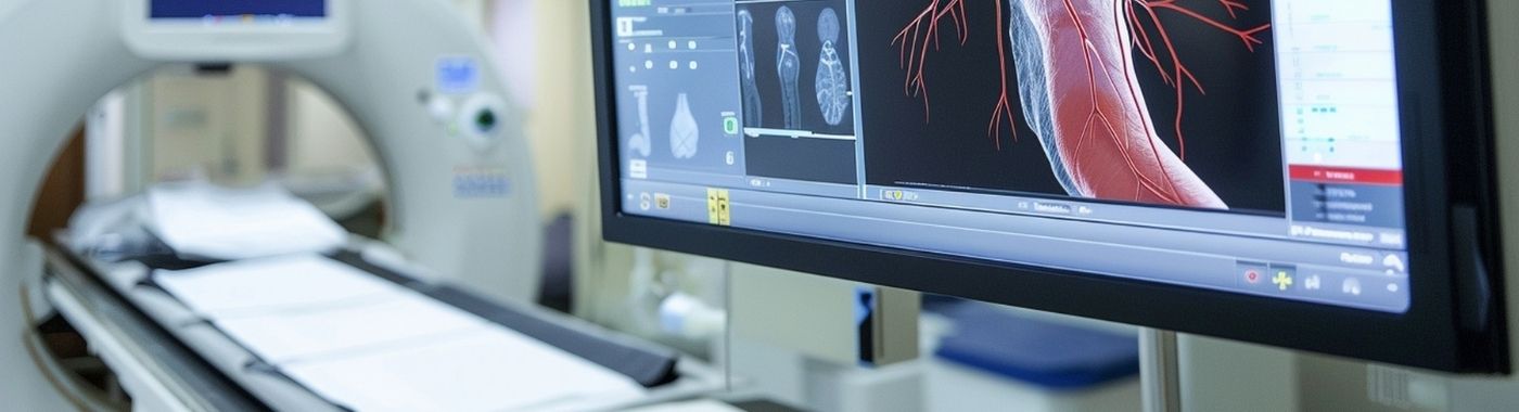
A coronary angiogram is an invasive X-ray test in which a thin catheter is passed into the heart arteries and iodinated contrast is injected to map narrowings, blockages and anatomy. The coronary angiogram guides decisions on medicines, angioplasty with stent, or bypass surgery. The angiogram shows artery lumen, not heart muscle damage or future heart attack risk.
Кључни одводи
- Намена: The coronary angiogram maps the inside of the coronary arteries more precisely than any non-invasive test.
- Ограничења: The angiogram shows the lumen only, it does not measure heart muscle function, viability, or predict when a plaque will rupture.
- Инвазиван: Unlike an echo test or a treadmill test, a coronary angiogram involves arterial puncture, contrast and radiation, so it carries a small but real risk.
- Not the same as angioplasty: Angiography is diagnostic; angioplasty (PTCA with stent) is treatment, sometimes performed in the same sitting.
- Тумачење: Findings must be read alongside symptoms, ECG, echo and functional tests by the cardiologist who requested the study. Results vary depending on individual clinical circumstances.
На први поглед
| Тачка | детаљ |
|---|---|
| Тип теста | Invasive catheter-based X-ray imaging of coronary arteries |
| Где је урађено | Catheterisation laboratory (cath lab) |
| Приступни пут | Usually radial artery at the wrist; sometimes femoral artery in the groin |
| Анестезија | Локални анестетик на месту убода, са или без благог седативног ефекта |
| Контраст | Iodinated contrast medium |
| Типично трајање поступка | About 20-45 minutes for a diagnostic study; longer if angioplasty follows |
| Total hospital time | Commonly a few hours to overnight, depending on access route, sedation and the unit's protocol |
| Зрачење | Yes, ionising radiation is used |
| Шта не може да уради | Cannot assess muscle viability, cannot exclude microvascular disease, cannot predict future events |
Такође познат као
- ЦАГ
- Cardiac catheterisation (angiography is one part of a cath study)
- Коронарна ангиографија
- "Heart ki angiography", "angio test", "dye test", "heart pipe check", terms commonly used by patients and families in India
- Left heart catheterisation with coronary angiography
What a coronary angiogram is
The coronary angiogram is a procedure in which a cardiologist punctures an artery, usually the radial artery at the wrist, and advances a fine, flexible catheter up to the origin of the coronary arteries. Iodinated contrast is injected and rapid X-ray images (cine runs) are recorded from several angles.
Because contrast fills the lumen (the channel through which blood flows), the images show where the channel is narrowed, irregular, or blocked, and how far disease extends. The angiogram also shows collateral vessels, bypass graft patency in patients who have had CABG, and anatomical variations such as anomalous coronary origin.
Coronary angiography is often combined with left ventricular pressure measurement, and increasingly with adjunctive tools: FFR/iFR (pressure wires to judge whether a moderate narrowing actually limits flow) and Интравенозни ултразвук/октаторна томографија (imaging catheters that look at the vessel wall itself). These add-ons exist precisely because the plain angiogram has limits.
Why a coronary angiogram is done
- Акутни коронарни синдром: In ST-elevation myocardial infarction, urgent angiography with primary angioplasty is the standard reperfusion strategy where a cath lab is available in time. In high-risk non-ST-elevation ACS, early invasive assessment is usual.
- Angina not controlled on medicines: To define anatomy and decide on stenting or bypass.
- Abnormal or high-risk non-invasive test: Strongly positive stress test, large ischaemia on stress echo or nuclear imaging, or severe stenosis suspected on CT coronary angiography.
- Before certain heart surgeries: For example, before valve surgery, to know whether coronary disease needs to be addressed at the same time.
- Unexplained heart muscle weakness: To check whether reduced pumping function is due to coronary disease.
- Follow-up of previous intervention: Recurrent symptoms after stenting or bypass.
Ко би требало да размотри овај тест
A coronary angiogram is not a screening test and is not appropriate for people without symptoms or risk assessment pointing towards significant coronary disease. It is generally considered when:
- Chest pain or breathlessness is typical of angina and persists despite optimal medical treatment.
- A heart attack is occurring or has recently occurred.
- A non-invasive test suggests a large area of the heart is at risk.
- Symptoms are strongly suggestive but non-invasive tests are inconclusive or not technically possible.
- Coronary anatomy must be known before valve or other cardiac surgery.
- There is unexplained left ventricular dysfunction, ventricular arrhythmia, or suspected coronary anomaly.
Обично је не the right first step for stable, low-risk chest pain, for reassurance without indication, or as a "package" health check. In those situations a clinical assessment, ECG, echo test and a functional or CT-based test are the reasonable starting points. Whether an angiogram is appropriate is a decision for your cardiologist.
What a coronary angiogram cannot detect or exclude
This is the part most often left out of patient information, and it matters.
- It does not show the artery wall. The angiogram outlines the lumen. Plaque that has remodelled outward can be extensive while the lumen still looks acceptable. A "normal" angiogram does not mean an absence of atherosclerosis.
- It cannot predict heart attacks. Many heart attacks arise from plaques that were not severely narrowing. A reassuring angiogram today does not guarantee freedom from future events, and risk factor treatment still matters.
- It does not reliably diagnose microvascular angina or coronary spasm. Angina with non-obstructive coronary arteries (ANOCA/INOCA) is common, especially in women. Diagnosing it needs additional testing such as provocation or coronary physiology studies, not the standard angiogram alone.
- It does not measure heart muscle function or viability. Echocardiography, cardiac MRI or nuclear perfusion imaging answer those questions.
- Visual severity estimates are subjective. Two experienced operators can grade the same moderate narrowing differently. For intermediate lesions, pressure-wire assessment is more informative than eyeballing the percentage.
- It says little about very small distal vessels, which may be poorly opacified or beyond resolution.
- It is not a valve, pericardium or aorta test. Those need echo or dedicated imaging.
A negative or normal-appearing angiogram describes what the images showed. It does not by itself exclude a cardiac cause of your symptoms. If symptoms persist or worsen despite a normal result, go back to your doctor rather than assuming the heart has been cleared.
Повезани, али различити прегледи
| Тест | Питање на које одговара | Кључна разлика |
|---|---|---|
| Coronary angiogram (invasive) | Exactly where and how severe are the luminal narrowings, and can they be treated? | Invasive; allows treatment in the same sitting; highest spatial detail of the lumen |
| ЦТ коронарна ангиографија (КТКА) | Are the coronary arteries free of significant disease in someone with low-to-intermediate likelihood? | Non-invasive; very useful for ruling out obstructive disease; heavy calcification, high or irregular heart rate and stents can degrade interpretation; cannot treat |
| Resting 2D echo (echo test) | How well is the heart pumping? Are the valves and pericardium normal? | Не приказује коронарне артерије; нормалан ехо не искључује коронарну артеријску болест |
| Treadmill test / stress ECG | Do symptoms and ECG changes appear with exertion? | Functional, not anatomical; limited in those who cannot exercise; both false positives and false negatives occur |
| Stress echo / nuclear perfusion (MPI) | Is any part of the heart muscle short of blood during stress, and how large is that area? | Shows functional significance and extent of ischaemia, not the precise anatomy |
| МРИ срца | Is the muscle scarred, inflamed or viable? | Best for tissue characterisation; not the standard test for coronary lumen anatomy |
| Coronary angioplasty (PTCA/PCI with stent) | Can the narrowing be opened and held open? | A treatment, not a diagnostic test; follows angiography |
| Peripheral / renal / cerebral angiography | Anatomy of arteries elsewhere in the body | Same technique, different territory, not interchangeable with coronary angiography |
None of these tests is universally superior. They answer different questions, and which comes first depends on how you present: a person with an evolving heart attack goes straight to invasive angiography, while someone with stable atypical chest pain and low risk is usually better served by a non-invasive test first.
Како припремити
Preparation instructions are set by the cath lab performing your procedure and vary between units. The written instructions given by your own team always take precedence over general information.
- Лекови: Bring a complete list, including blood thinners, antiplatelets, insulin and oral diabetes medicines, diuretics, metformin, herbal and AYUSH preparations, and gym supplements. Do not stop or change any prescribed medicine on your own, ask the prescriber or the cath lab team what to do on the day.
- Алергије: Report any previous reaction to iodinated contrast, and any significant asthma or drug allergy. Premedication protocols differ between units.
- Пост: Many units ask for a period of fasting before the procedure. The duration differs by unit and by whether sedation is planned.
- Основни тестови: Kidney function, haemoglobin, platelet count, coagulation tests, ECG and sometimes an echo are commonly obtained beforehand.
- Заштита бубрега: Adequate hydration before and after contrast is usual, but the type and volume of fluid must be individualised.
- Практични аранжмани: Arrange a companion to accompany you home. Do not drive yourself on the day. Remove jewellery and leave valuables at home; bring previous angiogram CDs, stent cards and discharge summaries.
Ако имате дијабетес на терапији за снижавање глукозе
Fasting while continuing the usual dose of insulin or a sulfonylurea can cause hypoglycaemia. Ask your prescriber specifically how to adjust doses on the morning of the procedure, and mention if you take metformin or an SGLT2 inhibitor, as units have specific protocols around contrast and these drugs.
Ако имате хроничну болест бубрега
Iodinated contrast carries a risk of contrast-associated kidney injury, higher with reduced eGFR, dehydration, diabetes and large contrast volumes. Tell the team your latest creatinine or eGFR. Contrast volume can often be minimised, and hydration planned carefully.
Ако имате срчану инсуфицијенцију или ограничење течности
Standard pre-procedure hydration advice can be unsafe if you have decompensated heart failure or a prescribed fluid limit. Fluid volumes must be set by your treating team, not by a generic instruction sheet.
У трудноћи
Coronary angiography uses ionising radiation and is undertaken in pregnancy only when the clinical need is compelling, such as a heart attack in pregnancy, with abdominal shielding and dose minimisation. Tell the team if you are or might be pregnant.
Код старијих одраслих
Frailty, low body weight, anaemia and reduced kidney reserve increase the risk of bleeding and contrast-related kidney injury. Radial access and lower contrast volumes are often preferred.
Код деце
Coronary angiography in children is uncommon and is generally performed for congenital coronary anomalies, Kawasaki disease sequelae or after cardiac transplantation, in paediatric cardiac centres and usually under general anaesthesia.
Шта се дешава током поступка
You lie awake on a narrow table with ECG leads, a blood pressure cuff and a pulse oximeter attached. An intravenous line is placed. The wrist or groin is cleaned and numbed with local anaesthetic. You may be given light sedation.
The cardiologist inserts a short sheath into the artery, then advances catheters to the heart. Contrast injection often produces a brief warm flush; some people feel a few seconds of chest discomfort or a fluttering sensation. You may be asked to hold your breath, cough, or keep still during image acquisition.
Most people describe pressure rather than pain. Tell the team immediately if you feel chest pain, severe discomfort, itching, breathlessness or nausea, these can be managed during the procedure.
At the end, the catheters are removed. For radial access a compression band is applied to the wrist; for femoral access manual pressure, a compression device or a closure device is used. Bed rest is usually required after femoral access, and much less after radial access.
Колико ми треба да стигнем до тамо
- Imaging itself: often 20-45 minutes for a straightforward diagnostic study.
- With angioplasty and stenting in the same sitting: considerably longer, depending on the complexity of the lesions.
- Посматрање након тога: a few hours after radial access in many units; longer, often with overnight stay, after femoral access, after intervention, or if you have other medical problems.
Exact timings are unit-specific and depend on scheduling, sedation, access route and your clinical condition.
Разумевање вашег извештаја
An angiogram report describes each major artery, left main, left anterior descending (LAD), left circumflex, right coronary artery, and their branches, with an estimate of narrowing as a percentage of the lumen diameter.
- Normal coronary arteries: No significant luminal narrowing was seen. This does not mean atherosclerosis is absent, nor that a cardiac cause of symptoms has been excluded.
- Mild / non-obstructive disease: Plaque is present but flow is not significantly limited. Usually managed medically with risk-factor treatment, not stenting.
- Moderate narrowing: Visual grading is least reliable here. Pressure-wire assessment (FFR/iFR) often decides whether treatment is needed.
- Severe narrowing: Flow-limiting disease that may be treated with angioplasty and stent or with bypass surgery, depending on the pattern, number of vessels, diabetes status, and pumping function.
- Тотална оклузија: The artery is completely blocked. Collateral vessels may be supplying the territory. Treatment depends on symptoms and whether the muscle beyond is viable.
- Други налази: Coronary ectasia or aneurysm, dissection, spasm, myocardial bridging, or anomalous origin. Each has its own management and should be explained to you specifically.
- Left ventricular function may be noted if ventriculography was performed.
Terms like "single vessel disease" or "triple vessel disease" describe how many major arteries are significantly involved. They describe anatomy, not prognosis on their own. The cardiologist who performed the study, with your symptoms and other tests in hand, is the person to interpret what the findings mean for you.
Када резултати могу бити обмањујући
Озбиљност може изгледати лажно већа
- Спазм изазван катетером, particularly of the right coronary artery, can mimic a narrowing; it resolves with intracoronary nitrate.
- Foreshortening and overlap of vessels in a single projection can exaggerate a narrowing, which is why multiple angles are taken.
- Low blood pressure or vasoconstriction during the study can make vessels appear smaller.
- Streaming of contrast or inadequate injection can create the appearance of filling defects.
Озбиљност може изгледати лажно мања
- Outward (positive) remodelling keeps the lumen looking normal despite a large plaque burden.
- Diffuse disease without a discrete narrow point may be under-read, because grading depends on comparing with a "normal" adjacent segment that may itself be diseased.
- Heavy calcification and dense overlapping structures can obscure the true lumen.
- Left main and ostial lesions can be missed if the catheter position obscures the segment.
- Small distal vessels and side branches may be inadequately opacified.
Technical and patient factors
- Movement, inability to hold breath, obesity and very high or irregular heart rates degrade image quality.
- Limits on contrast volume in kidney disease may reduce the number of views obtained.
- Previous bypass grafts can be difficult to engage and may be wrongly reported as occluded if not selectively imaged.
Ризици и безбедност
Coronary angiography is a well-established procedure performed in large numbers, and serious complications are uncommon. It is not risk-free, and the risk depends on your age, kidney function, the urgency of the procedure, the access route and whether treatment is performed at the same time. Results and risks vary depending on individual clinical circumstances.
Diagnostic angiography alone
- Common and usually minor: bruising, tenderness or a small haematoma at the puncture site; transient warmth or flushing from contrast; brief chest discomfort or arrhythmia during injection.
- Мање уобичајено: significant bleeding needing treatment, radial artery occlusion, pseudoaneurysm or arteriovenous fistula (more with femoral access), contrast-associated kidney injury, contrast hypersensitivity reactions, infection at the site.
- Rare but serious: heart attack, stroke, coronary dissection, serious arrhythmia, severe allergic reaction, and death. Risk is higher in emergency procedures, in unstable patients and in those with severe left main disease or advanced kidney disease.
- Зрачење: Dose is kept as low as reasonably achievable; cumulative dose matters if you undergo repeated procedures.
Angiography with angioplasty and stenting
Adding intervention increases procedural risk compared with diagnostic angiography alone, including a higher chance of vessel dissection, perforation, no-reflow, periprocedural myocardial injury, and larger contrast and radiation exposure. Stenting also commits you to a period of dual antiplatelet therapy, which increases bleeding risk. These are separate consent discussions.
Radial versus femoral access
Radial (wrist) access is generally associated with fewer access-site bleeding complications and earlier mobilisation than femoral access, which is why many units prefer it. Femoral access is still needed in some situations, for example when larger equipment is required, or when the radial route is unsuitable.
Red flags after a coronary angiogram
Emergency: go to the nearest emergency department or call for help now
- Chest pain, pressure or heaviness similar to or worse than your original symptoms, especially with sweating, nausea or breathlessness.
- Bleeding from the puncture site that does not stop with firm pressure, or rapid swelling in the groin or wrist.
- Sudden weakness or numbness of the face, arm or leg, difficulty speaking, or loss of vision.
- Fainting, severe breathlessness at rest, or palpitations with dizziness.
- A cold, pale, blue or pulseless hand or leg on the side of the puncture, or severe pain in that limb.
- Widespread rash with facial or throat swelling, wheeze or difficulty breathing after contrast.
Исти дан: контактирајте свој кардиолошки тим данас
- Expanding bruise or a lump at the site that is getting larger or increasingly painful.
- Грозница, ширење црвенила, топлина или исцедак на месту убода.
- New back or abdominal pain after femoral access.
- Markedly reduced urine output, or new swelling of the legs or face.
- Persistent vomiting preventing you from taking prescribed antiplatelet medicines.
Рутински преглед
- Stable, gradually fading bruising or mild tenderness at the site.
- Questions about your report, stent card, medicines or activity restrictions.
- Planning cardiac rehabilitation, return to work, or discussion of risk-factor treatment.
- Mild tingling or numbness at the wrist that is improving.
Посебне ситуације
Žene
Women more often have angina with non-obstructive coronary arteries. A "normal" angiogram in a woman with typical symptoms should prompt consideration of microvascular dysfunction or spasm rather than dismissal of the symptoms.
Дијабетес
Coronary disease in diabetes tends to be more diffuse and multi-vessel. This influences the choice between stenting and bypass surgery, and that decision is usually made by a heart team.
Хронична болест бубрега и дијализа
Contrast exposure needs careful planning. In people already on dialysis the concern shifts from kidney injury to fluid balance and timing of dialysis around the procedure.
Heart failure and severe valve disease
Lying flat may be difficult, and contrast load must be limited. Additional haemodynamic measurements may be made during the same study.
Prior bypass surgery
Graft angiography takes longer, uses more contrast and involves higher radiation than a first-time study. Bring your previous operation notes so the number and type of grafts are known.
Older adults and anticoagulated patients
Bleeding risk is the main consideration. The timing of anticoagulants around the procedure must be decided by the prescriber, not adjusted at home.
Coronary angiogram in the Indian context
- Earlier onset disease: Coronary artery disease is frequently seen at younger ages in South Asians than in many Western populations, and metabolic risk appears at lower body mass index in South Asians, so risk should not be dismissed on the basis of a "normal" weight.
- Реуматска болест срца: Valvular disease remains common in India. Coronary angiography is often performed before valve surgery in older patients or those with risk factors, and an echo test remains the primary valve assessment.
- Time to treatment: In heart attack, delays in reaching a cath-lab-capable hospital remain a major issue. If you have ongoing chest pain, call emergency services rather than arranging an outpatient appointment.
- Карактеристике анемије и таласемије: Iron-deficiency anaemia and haemoglobinopathy traits are common in India. Anaemia increases procedural and bleeding risk and can also cause breathlessness that mimics angina.
- Лекови и суплементи: Herbal, AYUSH and gym supplement use is common and often not reported. Disclose everything you take, as some agents affect bleeding and liver or kidney function.
- Друга мишљења: You are entitled to ask for the angiogram images and report, and to seek a second opinion before agreeing to stenting or bypass surgery, unless the situation is an emergency.
- Consent and data: Written informed consent is required, and your health data, including procedure images, is personal data handled under the Digital Personal Data Protection Act, 2023. Advertising claims of cures for heart disease are restricted under the Drugs and Magic Remedies (Objectionable Advertisements) Act, 1954.
Трошкови и осигурање у Индији
Costs differ widely between government, trust and private hospitals, between cities, and between a diagnostic study and a procedure that becomes therapeutic. Ask for a written estimate before admission.
Фактори који утичу на цену:
- Whether the procedure remains diagnostic, or angioplasty and stents are added in the same sitting.
- Access route, number of catheters and volume of contrast used.
- Use of adjunctive tools such as pressure wires, IVUS or OCT.
- Day-care versus overnight or ICU stay; room category.
- Pre-procedure investigations and cardiology consultation charges.
- Emergency out-of-hours procedures versus elective scheduling.
- City, hospital tier and whether the hospital is empanelled under a government scheme.
Тачке осигурања које треба проверити: whether your policy covers the procedure as a day-care or in-patient admission, waiting periods for pre-existing cardiac disease, room-rent capping and proportionate deduction clauses, whether stents and consumables are covered, and whether pre-authorisation is needed. Stent prices in India are subject to price ceilings notified by the National Pharmaceutical Pricing Authority; ask for the printed stent invoice and stent card. Government schemes such as Ayushman Bharat PM-JAY cover cardiac packages at empanelled hospitals for eligible beneficiaries.
Митови и чињенице
| Мит | Чињеница |
|---|---|
| "A normal angiogram means my heart is fine and I need no treatment." | A normal angiogram means no significant luminal narrowing was seen. Plaque, microvascular disease and other cardiac conditions can still be present, and risk-factor treatment still matters. |
| "An angiogram prevents heart attacks." | The angiogram is a diagnostic test. It informs treatment; it does not by itself reduce risk. Most heart attacks arise from plaques that were not severely narrowing. |
| "Angiogram and angioplasty are the same thing." | Angiography maps the arteries. Angioplasty with a stent is a treatment that may follow, sometimes immediately, sometimes after discussion. |
| "Every narrowing found needs a stent." | Mild and many moderate narrowings are best treated with medicines and risk-factor control. Some patterns of disease are better treated with bypass surgery. |
| "It is major open surgery." | Diagnostic angiography is a catheter procedure through a small puncture, usually under local anaesthetic. It is invasive but not open surgery. |
| "The dye will damage my kidneys permanently." | Contrast can affect kidney function, particularly with pre-existing kidney disease, dehydration or high contrast volumes. Most changes are transient, and risk can be reduced with planning. |
| "A CT angiogram can always replace an invasive angiogram." | CT coronary angiography is valuable, especially for excluding obstructive disease in lower-risk patients, but heavy calcification and stents limit it, and it cannot treat a blockage. |
| "If I am young and slim, my chest pain cannot be cardiac." | Coronary disease occurs at younger ages and at lower BMI in South Asians. Symptoms should be assessed, not assumed. |
Često postavljana pitanja
Is a coronary angiogram painful?
Most people feel a brief sting from the local anaesthetic and then pressure rather than pain. Contrast injection can cause a few seconds of warmth or mild chest discomfort. Significant pain is not expected, tell the team at once if you feel it, as it can be treated during the procedure.
Колико дуго ћу бити у болници?
The imaging usually takes 20-45 minutes for a diagnostic study. Observation afterwards is often a few hours with wrist access and longer, sometimes overnight, with groin access, after angioplasty, or if you have other medical conditions. Your unit will confirm the plan for you.
Can I eat and take my medicines before the procedure?
Fasting duration and medicine instructions are set by the performing unit and differ between hospitals. Do not stop or change any prescribed medicine yourself, particularly blood thinners, insulin or diabetes tablets. Ask your prescriber or the cath lab team exactly what to take on the day.
Will a stent be placed in the same sitting?
Sometimes. In an emergency such as a heart attack, treatment usually follows immediately. In stable cases the cardiologist may discuss findings with you or a heart team first. Consent for possible angioplasty is often taken in advance so treatment need not be delayed.
What is the difference between a coronary angiogram and a CT angiogram?
CT coronary angiography is non-invasive and useful for excluding significant blockages when the likelihood of disease is low to intermediate. Invasive coronary angiography gives finer detail of the lumen, allows pressure and imaging wire assessment, and permits treatment in the same sitting.
Is the radiation from an angiogram harmful?
The procedure uses ionising radiation, and dose is minimised using shielding, low-dose settings and limited screening time. For a single clinically indicated study the risk is small relative to the diagnostic benefit. Tell the team about previous cardiac procedures so cumulative exposure can be considered.
My angiogram was normal but chest pain continues. What next?
Persisting symptoms deserve reassessment, not dismissal. Possibilities include microvascular angina, coronary spasm, and non-cardiac causes such as acid reflux, musculoskeletal pain, anaemia or anxiety. Return to your cardiologist, who may arrange further cardiac testing or investigate other systems.
Могу ли да возим или да се вратим на посао после?
Do not drive on the day of the procedure if you received sedation. Many people resume light activity within a day or two after a diagnostic study, avoiding heavy lifting and strenuous use of the punctured limb for a few days. Timelines differ after angioplasty, follow your discharge advice.
How often can a coronary angiogram be repeated?
There is no fixed schedule. Repeat angiography is done when there is a clinical reason, such as recurrent or worsening symptoms, a new abnormal stress test, or planned staged intervention. Repeat studies are avoided without indication because of contrast and radiation exposure.
Извори
- 2021 ACC/AHA/SCAI Guideline for Coronary Artery Revasculari











 Најбоља болница у близини Ченаја


