Bronchoscopy is a procedure in which a pulmonologist passes a thin tube with a camera through the nose or mouth into the windpipe and airways. Bronchoscopy allows direct inspection of the airway lining, sampling of secretions and tissue, and certain treatments. Bronchoscopy does not examine lung tissue beyond the airways directly.
Key takeaways
- Bronchoscopy gives direct vision of the large and medium airways and allows washings, brushings and biopsies.
- Bronchoscopy does not replace a CT scan; imaging shows areas the scope cannot reach.
- A normal bronchoscopy does not exclude lung cancer, tuberculosis or interstitial lung disease.
- Flexible and rigid bronchoscopy are different procedures with different risks and anaesthesia needs.
- Preparation, fasting times and sedation practice differ between hospitals; the instructions from your own unit take precedence.
At a glance
| Feature | Detail |
|---|---|
| What it examines | Nose/throat passage, voice box, trachea, main and segmental bronchi |
| Main types | Flexible (diagnostic and therapeutic), rigid, EBUS (endobronchial ultrasound) |
| Anaesthesia | Local anaesthetic spray with or without conscious sedation; rigid bronchoscopy needs general anaesthesia |
| Typical procedure time | Diagnostic flexible bronchoscopy commonly 15-45 minutes; EBUS and therapeutic procedures often longer |
| Total time in hospital | Usually several hours, including consent, preparation and recovery monitoring |
| Admission | Most diagnostic flexible bronchoscopies are day-care; rigid and interventional cases may need admission |
| Results | Visual findings often discussed the same day; biopsy, cytology, culture and TB tests take longer |
Also known as
- Flexible fibreoptic bronchoscopy, FOB
- Lung camera test or "lung endoscopy" (common patient phrasing in India)
- Bronchoalveolar lavage (BAL), a sample taken during bronchoscopy, not a separate test
- EBUS-TBNA, endobronchial ultrasound-guided needle sampling of lymph nodes
- Hindi/vernacular usage: "phephde ki durbeen jaanch"
What bronchoscopy is
Bronchoscopy uses a bronchoscope, a flexible tube carrying a light, camera and a working channel. The bronchoscope is passed through a nostril or the mouth, past the vocal cords, into the trachea and then into the branching bronchi. Local anaesthetic is instilled along the way to reduce coughing.
Through the working channel, the pulmonologist can suction secretions, instil saline and collect it back (bronchoalveolar lavage), brush the airway lining, take forceps biopsies, pass a needle into adjacent lymph nodes, or deliver treatment. Rigid bronchoscopy uses a straight metal tube under general anaesthesia and is used mainly for airway obstruction, stenting, large foreign bodies and significant bleeding.
Why bronchoscopy is done
- Diagnostic sampling: suspected lung cancer, tuberculosis, fungal or atypical infection, especially when sputum tests are negative or the patient cannot produce sputum.
- Unexplained findings: a persistent shadow, mass, collapse or unresolving consolidation on chest X-ray or CT.
- Coughing up blood (haemoptysis): to look for a bleeding source in the airway.
- Persistent unexplained cough or hoarseness when other assessment has not explained it.
- Infection in immunocompromised patients: BAL samples for bacteria, mycobacteria, fungi, Pneumocystis and viruses.
- Therapeutic use: removing mucus plugs or inhaled foreign bodies, tumour debulking, stent placement, control of airway bleeding.
- Staging: EBUS-TBNA to sample mediastinal lymph nodes in known or suspected lung cancer.
Who should consider bronchoscopy
- Adults with an abnormal chest CT suggesting a central airway lesion or mass.
- People with suspected pulmonary tuberculosis who cannot produce sputum, or whose sputum tests are negative despite suggestive imaging, a common scenario in India.
- People coughing up blood repeatedly, particularly smokers and those over 40.
- People with recurrent pneumonia affecting the same area of the lung.
- Patients with reduced immunity (post-transplant, on chemotherapy, advanced HIV) and new lung infiltrates.
- Children with suspected inhaled foreign body, stridor or persistent lobar collapse, assessed by a paediatric pulmonologist.
Bronchoscopy is not a screening test. Bronchoscopy is not used for routine check-ups, breathlessness alone, or uncomplicated asthma or COPD.
What bronchoscopy cannot detect or exclude
- Peripheral lung lesions: nodules in the outer lung, away from an airway, are often beyond the reach and view of a standard bronchoscope. A negative bronchoscopy does not exclude a peripheral lung cancer. CT-guided biopsy, navigational bronchoscopy or surgical biopsy may be needed.
- Disease outside the airway wall: the bronchoscope sees the inside surface. Lymph nodes and masses beyond the wall need ultrasound guidance (EBUS) or needle sampling.
- Pleural disease: pleural effusion, empyema and pleural thickening are not assessed by bronchoscopy; these need pleural ultrasound, pleural fluid aspiration or thoracoscopy.
- Pulmonary embolism: bronchoscopy does not look at blood vessels. CT pulmonary angiography is the relevant test.
- Lung function: bronchoscopy says nothing about how well the lungs work. Spirometry and other pulmonary function tests measure that.
- Sampling error: a biopsy or lavage may miss the abnormal area even when disease is present. Culture and TB tests can be falsely negative, particularly in paucibacillary tuberculosis.
A "not identified" result means nothing abnormal was seen or grown in the samples taken. That is not the same as excluded. If symptoms continue despite a normal bronchoscopy, return to your doctor for reassessment.
Related but different examinations
| Examination | Question it answers | Key difference |
|---|---|---|
| Flexible bronchoscopy | What does the airway lining look like, and can I sample it? | Awake or lightly sedated; limited to airway reach |
| Rigid bronchoscopy | Can the airway be opened, secured or cleared? | General anaesthesia; better for bleeding, stents, large foreign bodies |
| EBUS-TBNA | Are the mediastinal lymph nodes involved? | Ultrasound on the scope tip allows needle sampling beyond the airway wall |
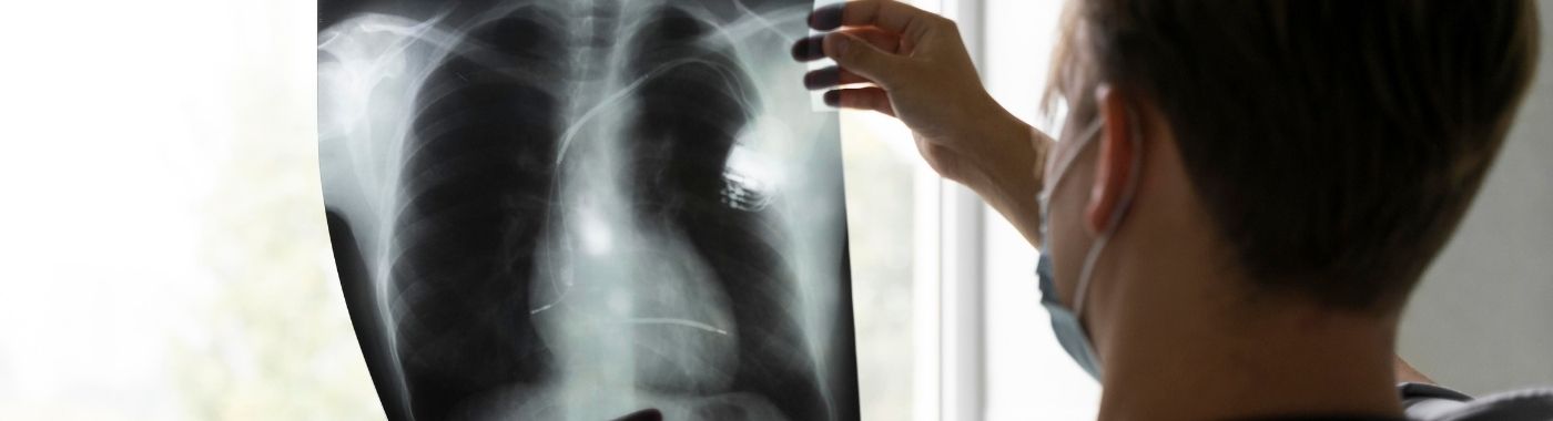
| Chest X-ray | Is there an obvious abnormality? | Quick, low dose, but insensitive to small or central lesions |
| CT chest | Where exactly is the abnormality, and how extensive? | Maps the whole lung including peripheral areas; no tissue obtained |
| CT-guided lung biopsy | What is a peripheral lesion made of? | Needle through the chest wall; higher pneumothorax risk than bronchoscopy |
| Sputum tests (including molecular TB testing) | Is there infection I can detect non-invasively? | No procedure needed; should usually be tried first for suspected TB |
| Thoracoscopy / pleuroscopy | What is happening in the pleural space? | Examines the lung surface and pleura, not the airways |
Which test comes first depends on the clinical problem, not on a ranking. For suspected TB with productive cough, sputum testing usually comes before bronchoscopy. For a peripheral nodule, CT and a CT-guided approach are often more appropriate. For a central mass or haemoptysis, bronchoscopy is frequently the more direct route.
How to prepare
Preparation protocols are unit-specific. Fasting duration, sedation policy, drug-hold advice and escort requirements vary between hospitals. Follow the written instructions given by the unit performing your bronchoscopy; those instructions take precedence over anything on this page.
- Fasting: most units ask for a period of nil by mouth before sedation. Your unit will tell you the exact timing for food and clear fluids.
- Medicines: bring a full list, including inhalers, herbal and AYUSH preparations and supplements. Do not stop or change any prescribed medicine yourself, speak to the prescriber or the bronchoscopy unit.
- Blood thinners and antiplatelets: decisions about warfarin, newer oral anticoagulants, clopidogrel or aspirin depend on why you take them and whether a biopsy is planned. This must be decided by your treating doctor.
- Tests before the procedure: units commonly check recent imaging, oxygen levels and sometimes blood counts and clotting.
- Consent: risks, benefits and alternatives are discussed before you sign.
- Escort and travel: if sedation is used, arrange for someone to accompany you home. Do not drive, operate machinery or sign legal documents for the rest of the day.
- Dentures, loose teeth, jewellery are usually removed before the procedure.
Diabetes on glucose-lowering treatment
Fasting with insulin or sulfonylureas can cause hypoglycaemia. Tell the unit in advance, ask for an early slot where possible, and get specific instructions from your diabetes prescriber about doses on the day. Carry a glucose source.
Heart failure, advanced CKD or fluid restriction
Standard advice about drinking clear fluids may conflict with a prescribed fluid restriction. Tell the unit about your restriction and follow the individualised plan given.
Pregnancy
Bronchoscopy in pregnancy is uncommon and reserved for situations where the information is needed urgently. Sedation choices, positioning and monitoring are altered, and the obstetric team is involved. Tell the unit if you are or might be pregnant.
Children
Paediatric bronchoscopy uses smaller scopes and is usually done under general anaesthesia by teams with paediatric airway experience. Fasting times for children differ from adults and are set by the anaesthetist.
Older adults and people with significant heart or lung disease
Reduced respiratory reserve, unstable angina, recent heart attack, uncontrolled arrhythmia or severe hypoxaemia all increase risk and may mean the procedure is deferred, modified, or done in a monitored setting.
What happens during bronchoscopy
- Setting up: you lie on a couch or sit semi-reclined. A clip monitors oxygen saturation; ECG and blood pressure are usually monitored. Oxygen is given through the nose or mouth.
- Numbing: local anaesthetic is sprayed or gargled to numb the nose and throat. This tastes bitter and makes the throat feel swollen, that sensation is expected.
- Sedation: if used, sedation is given through a vein. Most people are drowsy and remember little, but bronchoscopy is generally not done under full general anaesthesia unless the case requires it.
- The examination: the scope passes through a nostril or the mouth, past the vocal cords, into the airways. Coughing is common and is managed with extra local anaesthetic. You can breathe throughout, the scope is much narrower than the windpipe.
- Sampling: lavage, brushings, forceps biopsy or needle sampling as planned. Biopsies themselves are not usually felt as pain, as the airway lining has no pain fibres of the kind found in skin.
- Afterwards: you are observed until sedation wears off and oxygen levels are stable. Eating and drinking are withheld until the throat numbness has fully resolved, to reduce the risk of choking.
How long it takes
A straightforward diagnostic flexible bronchoscopy commonly takes 15-45 minutes of scope time. EBUS, multiple biopsies or therapeutic work such as stenting or foreign body removal can take considerably longer. Including registration, consent, preparation and recovery, plan on being at the hospital for several hours. Timings vary with case complexity and unit workflow.
Understanding your report
Bronchoscopy does not produce a number or a reference range. The report describes what was seen and what was sampled.
- Visual findings: normal airway anatomy, inflammation, secretions, narrowing, external compression, a growth, or active bleeding, with the exact segment named.
- Procedures performed: lavage, brushing, biopsy sites, needle passes, therapeutic steps.
- Laboratory results that follow separately: histopathology, cytology, bacterial and fungal culture, molecular TB testing and mycobacterial culture. Mycobacterial culture in particular takes weeks.
"Normal bronchoscopy" means the visible airways looked normal at the time of the examination. It does not mean the lungs are disease-free. The final meaning of the findings comes from your pulmonologist, who combines the report with your symptoms, imaging and laboratory results. Results vary depending on individual clinical circumstances.
When results can be misleading
Falsely reassuring (disease present but not found)
- Peripheral lesions out of reach of the scope.
- Biopsy taken from the edge of a lesion, returning only inflammation or necrosis.
- Paucibacillary tuberculosis with negative smear, negative molecular test and even negative culture.
- Antibiotics or anti-tubercular therapy started before sampling, reducing culture yield.
- Small lavage volume or dilute sample lowering the concentration of organisms.
Falsely alarming (abnormality suggested where none exists)
- Trauma from the scope or suction causing redness or bleeding that can mimic inflammation.
- Contamination of samples with upper airway organisms, leading to growth of bacteria that are not causing disease.
- Colonising fungi such as Candida or Aspergillus grown from lavage in people who do not have invasive infection.
- Crush artefact in small biopsies making cells look atypical.
Discordant results, for example a suspicious CT with a negative biopsy, usually mean further sampling or follow-up imaging rather than closure of the issue.
Risks and safety
Bronchoscopy is generally well tolerated, but no procedure is risk-free, and the risk profile differs by type. Risk is higher with biopsies, in people with low oxygen levels, bleeding tendencies, pulmonary hypertension or significant heart disease.
Diagnostic flexible bronchoscopy
Common and usually self-limiting: sore throat, hoarseness, cough, small amounts of blood-streaked sputum, transient fall in oxygen saturation, low-grade fever for a few hours after lavage. Less common: bronchospasm, arrhythmia, reaction to local anaesthetic or sedation.
Bronchoscopy with biopsy
Bleeding risk rises with endobronchial and transbronchial biopsy. Transbronchial lung biopsy carries a recognised risk of pneumothorax (collapsed lung), which may need a chest drain. Units often arrange a post-procedure chest X-ray or ultrasound when transbronchial biopsy is performed.
EBUS-TBNA
Generally low complication rates. Recognised issues include bleeding at the needle site, infection of a sampled node or cyst, and longer sedation exposure.
Rigid and interventional bronchoscopy
Requires general anaesthesia, with the associated anaesthetic risks. Additional risks include dental or airway injury, airway perforation, and complications related to stents, laser or cryotherapy. These procedures are performed in centres with the appropriate surgical and anaesthetic backup.
Red flags after bronchoscopy
Emergency: go to the nearest emergency department now
- Coughing up more than a small amount of fresh blood, or blood that keeps increasing.
- Sudden severe breathlessness or sharp one-sided chest pain, which may indicate pneumothorax.
- Bluish lips or fingertips, confusion, or collapse.
- Noisy breathing, stridor or a feeling of the throat closing.
Same day: contact the bronchoscopy unit or your doctor today
- Fever above 38癈 persisting beyond the first several hours, or with shaking chills.
- Ongoing blood-streaked sputum beyond 24 hours.
- Chest discomfort that is new and does not settle.
- Vomiting or choking on attempting to eat or drink after the procedure.
Routine appointment
- Mild sore throat or hoarseness lasting a few days.
- Tiredness for a day after sedation.
- Questions about pending biopsy or culture results, or about next steps.
Special situations
Pregnancy
Bronchoscopy is performed in pregnancy only when the result will change urgent management. Positioning, oxygen monitoring and drug choice are adjusted, and the obstetric team is involved in planning.
Children
Children are usually assessed for inhaled foreign body, stridor, persistent collapse or recurrent infection. Paediatric flexible and rigid bronchoscopy are performed under anaesthesia by teams trained in paediatric airways.
Older adults
Sedation doses are typically reduced and monitoring is closer. Cognitive effects of sedation can last longer; arrange company for the day.
People with diabetes on medication
Fasting requires an individualised plan for insulin and oral glucose-lowering drugs, arranged with the prescriber before the appointment.
Advanced kidney disease or heart failure
Fluid, sedation and oxygen management need individual planning. Tell the unit about dialysis schedules and fluid restrictions.
Reduced immunity
Bronchoscopy with lavage is frequently the most useful way to identify the organism causing lung infiltrates in transplant recipients, people on chemotherapy and those with advanced HIV. A broad panel of tests is sent from a single lavage.
Bronchoscopy in the Indian context
- Tuberculosis: India carries a large share of the world's TB burden. Bronchoscopy with lavage is widely used when sputum cannot be produced or is repeatedly negative despite suggestive imaging. Molecular TB tests and mycobacterial culture with drug-susceptibility testing are commonly sent on lavage samples, in line with national TB programme practice.
- Non-resolving pneumonia and lung cancer: in smokers, bidi users and those with heavy biomass or air-pollution exposure, a persistent shadow may need bronchoscopic sampling rather than repeated courses of antibiotics.
- Foreign body inhalation: aspirated groundnut, chana and other food particles in toddlers are a recognised paediatric emergency indication for rigid bronchoscopy.
- Anti-tubercular therapy: tell the team if you are on ATT, as treatment already started can affect sample yield and interpretation.
- Infection control: reprocessing and disinfection of bronchoscopes follows defined hospital protocols; you may ask the unit about theirs.
- Access: flexible bronchoscopy is available in most tertiary centres; EBUS, navigational bronchoscopy and interventional rigid bronchoscopy are concentrated in larger hospitals.
Cost and insurance in India
Costs differ widely between government, trust and private hospitals, and between cities. Prices quoted online are indicative only; ask the performing unit for a written estimate for your specific procedure.
Factors that affect the cost:
- Type of procedure, diagnostic flexible bronchoscopy, EBUS-TBNA, rigid or interventional bronchoscopy.
- Anaesthesia: topical only, conscious sedation, or general anaesthesia with an anaesthetist.
- Day-care versus inpatient admission, and duration of recovery monitoring.
- Number and type of samples, and the laboratory tests requested, histopathology, cytology, molecular TB testing, mycobacterial and fungal culture, special stains.
- Consumables such as biopsy forceps, needles, cryoprobes, stents.
- Pre-procedure investigations and post-procedure imaging.
- City, hospital category and doctor's fees.
Insurance: bronchoscopy performed during an admission is usually covered under an indemnity health policy, subject to waiting periods and policy terms. Day-care procedures are covered by many policies but not all. Cover under government schemes such as Ayushman Bharat PM-JAY depends on the empanelled package and hospital. Confirm coverage and pre-authorisation with the hospital's insurance desk before the date.
Myths and facts
| Myth | Fact |
|---|---|
| Bronchoscopy will block my breathing | The bronchoscope is far narrower than the trachea, and oxygen is supplied throughout. Breathing continues during the examination. |
| Bronchoscopy is very painful | Most people describe pressure, a numb throat and coughing rather than pain. Local anaesthetic and, in many units, sedation are used. |
| A normal bronchoscopy means my lungs are fine | Bronchoscopy sees only the airways it can reach. Peripheral lesions, pleural disease and clots are not assessed. |
| A CT scan can always replace bronchoscopy | CT maps the lung but cannot obtain tissue or fluid. Diagnosis of cancer and infection usually requires a sample. |
| Bronchoscopy spreads cancer | There is no credible evidence that bronchoscopic sampling spreads lung cancer. Delayed diagnosis is the greater harm. |
| Bronchoscopy is only for cancer | Infection, particularly tuberculosis, is one of the commonest reasons for bronchoscopy in India. |
Frequently asked questions
How long does a bronchoscopy take?
Scope time for a routine diagnostic flexible bronchoscopy is commonly 15-45 minutes. EBUS, multiple biopsies or therapeutic procedures take longer. Allow several hours at the hospital overall, covering consent, preparation, the procedure and recovery from sedation. Exact timings depend on the case and the unit's workflow.
Will I be asleep during bronchoscopy?
Most diagnostic flexible bronchoscopies use local anaesthetic to the throat, often with conscious sedation, so you are drowsy but breathing on your own. Rigid and most paediatric bronchoscopies need general anaesthesia. Your unit will explain which approach applies and why before you consent.
When will I get my bronchoscopy results?
The pulmonologist can usually describe what was seen soon after the procedure. Laboratory results arrive separately: histopathology and cytology take days, molecular TB tests are quicker, and mycobacterial culture takes several weeks. Your treating doctor interprets all of these together with your imaging and symptoms.
Can bronchoscopy detect lung cancer reliably?
Bronchoscopy diagnoses many central lung cancers by direct biopsy. Peripheral tumours away from an airway are frequently missed, so a negative bronchoscopy does not exclude lung cancer. If suspicion remains, CT-guided biopsy, navigational bronchoscopy or surgical sampling may be recommended.
Is bronchoscopy better than a CT chest?
The two answer different questions. CT maps where an abnormality is, including areas a scope cannot reach, but provides no tissue. Bronchoscopy provides direct vision and samples but covers a limited territory. They are usually complementary, with CT typically done first to guide sampling.
Can I eat immediately after bronchoscopy?
No. The throat stays numb for a period after local anaesthetic, and swallowing while numb risks choking or aspiration. Your unit will tell you when to try sips of water first, usually after checking that your cough and swallow reflexes have returned.
Do I need to stop my blood thinners before bronchoscopy?
That decision belongs to your prescriber and the bronchoscopy team. It depends on the drug, why you take it, and whether a biopsy is planned. Do not stop any anticoagulant or antiplatelet medicine on your own, contact the prescribing doctor well before the appointment.
Is bronchoscopy safe if I already have lung disease?
Bronchoscopy is often performed in people with lung disease, but existing disease can raise risk. Low oxygen levels, severe COPD, pulmonary hypertension or unstable heart disease may lead the team to modify, defer or monitor the procedure more closely. Risk is assessed individually before proceeding.
Can children have a bronchoscopy?
Yes. Paediatric bronchoscopy is used for suspected inhaled foreign bodies, stridor, persistent lung collapse and recurrent infection. Smaller scopes are used, almost always under general anaesthesia, by teams experienced in paediatric airways. Fasting instructions for children are set by the anaesthetist and differ from adult guidance.
Sources
- British Thoracic Society guideline on flexible bronchoscopy in adults.
- American Thoracic Society and European Respiratory Society statements on bronchoscopic sampling and interventional pulmonology.
- National Tuberculosis Elimination Programme (India) technical guidance on diagnosis of pulmonary tuberculosis.
- World Health Organization Global Tuberculosis Report.
- Indian Chest Society / National College of Chest Physicians guidance on bronchoscopy practice.