


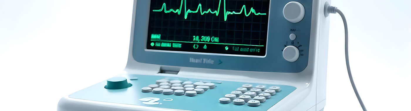
An ECG (electrocardiogram, commonly called an "ECG test" or "heart tracing") records the heart's electrical activity through electrodes placed on the chest and limbs. The ECG helps identify rhythm disturbances, conduction block, chamber strain and patterns suggesting a heart attack. A normal ECG does not exclude coronary artery disease or intermittent arrhythmia.
Klíčové jídlo s sebou
- Fast and non-invasive: a standard 12-lead ECG records for a few seconds and causes no pain.
- It is a snapshot: a normal ECG describes the heart's electrical activity only during those seconds.
- It does not image arteries or valves: an ECG shows electrical signals, not blood flow, blockages or valve structure.
- Interpretace potřebuje kontext: the same tracing can be normal in one person and significant in another.
- Persistent symptoms override a normal result: if chest pain or palpitations continue, return to your doctor.
Na první pohled
| vlastnost | Detail |
|---|---|
| Co je zaznamenáno | Electrical activity of the heart from 10 electrodes, displayed as 12 leads |
| Záření | None; the machine records signals, it does not send current into the body |
| PŘÍPRAVA | Obvykle žádné; instrukce jednotky mají přednost |
| Doba záznamu | A few seconds of tracing; typically 5-10 minutes in the room including electrode placement |
| Nepohodlí | Cool gel or adhesive pads; some chest hair may need trimming |
| Hlavní silné stránky | Rhythm, conduction, acute ischaemia patterns, electrolyte and drug effects |
| Hlavní slepá místa | Intermittent arrhythmia, stable coronary disease, valve and pump function |
Také známý jako
- ECG, EKG (German-derived spelling, elektrokardiogramm)
- Electrocardiogram, electrocardiography
- "ECG test", "heart tracing", "heart graph test", "12-lead ECG"
- Hindi/vernacular usage in Indian clinics: "dil ka ECG", "dil ki jaanch wali machine"
What an ECG is
Each heartbeat begins as an electrical impulse in the sinus node, spreads through the atria, passes the atrioventricular node and travels into the ventricles. The ECG picks up these voltage changes at the skin surface and prints them as waves: the P wave (atrial activation), the QRS complex (ventricular activation) and the T wave (ventricular recovery). Ten electrodes generate twelve different electrical "views" of the heart.
The ECG machine is a listening device. It does not pass current into the body.
Why an ECG is done
- Chest pain or chest pressure: to look for patterns of acute ischaemia or infarction, and to guide immediate treatment decisions.
- Palpitations, blackouts or near-fainting: to document rhythm if the symptom is present, and to look for underlying conduction or repolarisation abnormalities.
- Dušnost: as one part of a wider cardiac and respiratory assessment.
- Known heart disease: follow-up of arrhythmia, previous myocardial infarction, heart failure or cardiomyopathy.
- Pacemaker or ICD checks: spolu s formálním výslechem zařízení.
- Medication and electrolyte monitoring: some drugs affect the QT interval; potassium and calcium disturbances change wave shapes.
- Before surgery or before starting certain treatments, where the clinical situation warrants it.
Kdo by měl podstoupit tento test
- Anyone with acute chest pain, pressure, or pain spreading to jaw, neck or arm: this needs an emergency ECG, not a planned appointment.
- People with palpitations, unexplained fainting, or documented irregular pulse.
- People with known coronary artery disease, heart failure, valve disease or rheumatic heart disease as directed by their cardiologist.
- People with hypertension, diabetes or chronic kidney disease when a doctor is assessing cardiac involvement.
- People on medicines known to affect cardiac conduction or the QT interval, when the prescriber advises monitoring.
- Athletes and competitive sportspersons where a screening protocol applies, and people with a family history of sudden cardiac death or inherited heart disease.
- People being assessed before major surgery, based on age, symptoms and comorbidity rather than routinely for everyone.
A routine ECG in a person with no symptoms and no risk factors often adds little and can generate incidental findings that lead to further testing. Whether the ECG is useful for you is a clinical judgement.
What an ECG cannot detect or exclude
Toto je nejvíce nepochopená část testu.
- It cannot exclude coronary artery disease. A resting ECG is frequently normal in people with significant narrowing of the coronary arteries. The ECG does not visualise the coronary arteries at all.
- It cannot exclude a heart attack on its own. Early in an infarction the ECG may look normal or near-normal. Diagnosis usually requires serial ECGs plus cardiac troponin and clinical assessment.
- It cannot reliably capture intermittent arrhythmia. If the abnormal rhythm is not happening during those few seconds, the tracing will look normal. Ambulatory monitoring is needed.
- It does not measure pump function. Ejection fraction, valve narrowing or leak, pericardial fluid and clots are echocardiography questions, not ECG questions.
- It does not measure blood pressure, cholesterol or blockage percentage.
- It cannot diagnose lung or aortic emergencies. Pulmonary embolism and aortic dissection may produce non-specific ECG changes or none; imaging is required.
- "ECG normal" means no abnormality was identified in that recording. It is not the same as "heart disease excluded". If your symptoms persist or return despite a normal ECG, go back to your doctor.
Související, ale odlišná vyšetření
| test | Otázka to odpovídá | Co to nedělá |
|---|---|---|
| Resting 12-lead ECG | What is the electrical activity right now? | Does not show arteries, valves or pump function; misses intermittent rhythm problems |
| Stress (exercise) ECG / TMT | Objevují se změny nebo příznaky EKG při námaze? | Not suitable if the person cannot exercise; false positives and false negatives both occur, and results are less reliable when the resting ECG is already abnormal |
| Holter monitor (24-48 hours or longer) | What rhythms occur over a day or more of ordinary life? | Misses events that fall outside the recording window |
| Event / patch monitor, implantable loop recorder | What is the rhythm during infrequent symptoms? | Depends on symptoms occurring and, for some devices, on the patient activating them |
| Echokardiogram („echo test“, 2D echo) | Structure and function, chamber size, valves, ejection fraction, pericardium | Does not show the electrical rhythm in detail; a resting echo does not visualise coronary arteries |
| Cardiac troponin (blood test) | Došlo k poranění srdečního svalu? | Does not localise the problem or show rhythm; can rise in non-cardiac illness and in kidney disease |
| CT coronary angiography / invasive angiography | Jsou koronární tepny zúžené a kde? | Does not assess rhythm; involves radiation and contrast, with their own risks |
| Smartwatch or single-lead handheld ECG | Screening prompt for possible atrial fibrillation | One or few leads only; cannot substitute for a diagnostic 12-lead ECG or assess ischaemia |
These tests answer different questions. Which one comes first depends on the presentation: sudden severe chest pain leads to an immediate ECG and troponin; episodic palpitations lead towards ambulatory monitoring; breathlessness with a murmur leads towards echocardiography; exertional chest pain in a stable patient may lead to stress testing or CT coronary angiography depending on risk and local availability.
Jak se připravit
Most units require no special preparation for a resting ECG. Protocols differ between hospitals and diagnostic centres, and the instructions given by your own unit take precedence over anything written here.
- Žádný půst is needed for a standard resting ECG.
- Oblečení: wear something that allows access to the chest, wrists and ankles. Two-piece clothing is easier than a one-piece garment.
- Kůže: avoid oil, moisturiser or talcum powder on the chest that day, as these weaken electrode contact. Some men may need small patches of chest hair trimmed.
- Léky: bring your current list. Do not stop, skip or change any prescribed medicine before an ECG unless the prescribing doctor has told you to.
- Heavy exertion and stimulants: vigorous exercise, a large amount of caffeine, tobacco or alcohol shortly before the test can raise the heart rate and make interpretation harder. Rest for a few minutes before the recording.
- Řekněte to personálu if you have a pacemaker or ICD, a skin condition or adhesive allergy, chest dressings, or if you are in pain.
Pokud jste těhotná
A resting ECG is used in pregnancy when clinically indicated. There is no radiation. Heart rate is normally higher in pregnancy and some axis and repolarisation changes occur, so the tracing is read against pregnancy norms. Lying flat may be uncomfortable in later pregnancy, tell the technician, as a tilted position may be used.
Děti
Paediatric ECGs use smaller electrodes and, importantly, age-specific interpretation. Heart rates, intervals and wave patterns that are normal in a newborn would be abnormal in an adult. A paediatric ECG should be reported against paediatric norms.
Starší dospělí
Minor conduction changes and non-specific abnormalities are common with age and are not automatically disease. Comparison with an earlier ECG is often more informative than the tracing alone.
Lidé s diabetem, chronickým onemocněním ledvin nebo srdečním selháním
No fluid or fasting restriction applies to a resting ECG, so instructions for these groups are unchanged. Symptoms of ischaemia may be blunted or atypical in long-standing diabetes, so an ECG can be requested for vaguer symptoms such as unusual fatigue or breathlessness. In CKD and heart failure, potassium and other electrolyte shifts can alter the ECG independently of the heart itself.
Pacemaker or implanted device
An ECG can be recorded safely with a pacemaker or ICD in place, and helps show whether the device is pacing. It does not replace formal device interrogation by the cardiology team.
What happens during an ECG
- Umístění: you lie on a couch. The chest, wrists and ankles are exposed as needed; a sheet or gown is used for privacy, and you may request a female attendant.
- Umístění elektrody: ten electrodes are attached with gel or adhesive pads, six on the chest, one on each limb. Accurate placement matters, because misplaced leads produce misleading tracings.
- Záznam: you are asked to lie still, breathe normally and not talk for a few seconds. Movement, shivering and talking create artefact.
- Po: the electrodes are peeled off. There is no recovery time and you can return to normal activity at once. Gel can be wiped off; the skin may be pink briefly where pads were attached.
Jak dlouho proces trvá
- The tracing itself: usually around 10 seconds of recording.
- V místnosti: commonly 5-10 minutes including positioning and electrode placement.
- Whole visit: often 15-20 minutes with registration and waiting, though this depends entirely on the unit.
- Zpráva: in an emergency the ECG is read within minutes at the bedside. For outpatient tests, reporting time depends on the unit's workflow, ask at the reception when collecting.
Understanding your ECG report
An ECG report usually has three parts: measured numbers, a computer-generated interpretation, and the doctor's own reading. The doctor's reading is what counts. Automated interpretations are frequently wrong, and over-call abnormalities in particular.
Terms you may see:
- Normal sinus rhythm: the impulse is starting in the normal place and travelling normally.
- Sinus tachycardia / sinus bradycardia: a normal rhythm that is faster or slower than usual. Both have many innocent causes, anxiety, fever, pain, dehydration, anaemia, thyroid disease, fitness level or medication.
- Fibrilace síní: an irregular rhythm from the atria, important because of stroke risk and because it often needs specific treatment.
- Ectopics / premature beats: extra beats. Often benign, but frequent ectopy or ectopy with symptoms is assessed further.
- First-degree AV block, right bundle branch block, incomplete RBBB: conduction patterns that may be a normal variant in some people and significant in others.
- Left bundle branch block, new ST elevation or depression, T-wave inversion, Q waves: patterns that need prompt clinical correlation and comparison with any earlier ECG.
- Left ventricular hypertrophy "by voltage criteria": an electrical suggestion of thickened muscle, not a measurement. Echocardiography assesses this properly.
- Prolonged or short QT: relevant to arrhythmia risk and to certain medicines. Discuss with the prescriber, and do not stop a drug on your own.
- "Non-specific ST-T changes": a very common phrase that means the tracing is not textbook-normal but is not pointing to any one diagnosis. On its own it is rarely a cause for alarm; in someone with symptoms it prompts further assessment.
Deliberately, no numeric reference intervals are listed on this page. Normal ranges for heart rate and for the PR, QRS and QT intervals depend on age, sex, heart rate, body habitus, medication and the measurement method used, and are applied by the reporting doctor. A single number outside a textbook range is not a diagnosis. Results vary depending on individual clinical circumstances, and your ECG must be interpreted in context by the doctor who requested it.
When ECG results can be misleading
Nálezy, které mohou vypadat falešně abnormálně
- Lead misplacement or reversed limb leads: can mimic infarction, axis deviation or rhythm abnormality.
- Movement, shivering, tremor or talking: artefact can imitate arrhythmia. Parkinsonian tremor and electrical interference are classic culprits.
- Anxiety, pain, fever, recent exertion or stimulants: raise the rate and may cause ST-T changes.
- Body habitus, breast tissue, emphysema or large body size: alter signal amplitude and can falsely suggest, or mask, hypertrophy.
- Normální varianty: early repolarisation and juvenile T-wave patterns in young, fit or athletic people are often over-read as ischaemia.
- Computer interpretation: algorithms tend to over-report abnormality, and a printed word like "infarct" on the strip is not a diagnosis.
Findings that can look falsely normal or falsely reassuring
- Very early myocardial infarction: the ECG can be unremarkable in the first hours; repeat tracings often change.
- Stable coronary disease: commonly produces a completely normal resting ECG.
- Intermittent arrhythmia: invisible unless it happens during the recording.
- Pre-existing left bundle branch block, paced rhythm or old infarction: can mask new ischaemic changes.
- Posterior or isolated territory infarction: may be poorly represented on standard leads.
- Účinky léků: some drugs slow the rate or alter conduction, softening findings.
Because of both directions of error, the ECG is read alongside your symptoms, examination, previous tracings and, where needed, blood tests and imaging.
Rizika a bezpečnost
Resting 12-lead ECG
Recording a resting ECG is a very low-risk procedure. There is no radiation, no needle and no current delivered to the body. The realistic issues are minor skin irritation or redness from adhesive pads, discomfort if chest hair is trimmed, and a brief loss of privacy during electrode placement.
Ambulatory monitoring (Holter, patch, event monitor)
The main problems are skin irritation under long-worn electrodes, itching, and restrictions on bathing while the device is attached. Follow the unit's instructions for the specific device.
Stress (exercise) ECG
A stress ECG carries a different and higher risk profile than a resting ECG, because the heart is deliberately stressed. Uncommon but recognised risks include provoked chest pain, arrhythmia, marked blood pressure change, fainting and, rarely, serious cardiac events. It is performed with supervision and resuscitation facilities available, and it is not appropriate for everyone, for example, during suspected acute coronary syndrome or in some forms of severe valve disease. The doctor requesting it weighs these factors individually.
Safety statements for one type of ECG should not be applied to the whole family of tests.
červené vlajky
Emergency: go to the nearest emergency department now, or call emergency services
- Chest pain, pressure, heaviness or burning lasting more than a few minutes, especially with sweating, nausea, breathlessness, or pain spreading to the jaw, neck, back or arm.
- Fainting or collapse, particularly during exertion or without warning.
- Sudden severe breathlessness, or breathlessness that stops you lying flat.
- Palpitations with chest pain, faintness, confusion or collapse.
- Sudden weakness of one side, facial droop or slurred speech (possible stroke, which can accompany atrial fibrillation).
- Very slow or very fast pulse with dizziness or feeling about to pass out.
Do not wait for an outpatient ECG appointment in these situations, and do not drive yourself.
Tentýž den: kontaktujte svého lékaře ještě dnes
- New or worsening chest discomfort on exertion that settles with rest.
- Palpitations that are persistent, or a newly irregular pulse.
- Near-fainting, unexplained falls, or new marked breathlessness on mild activity.
- New swelling of both ankles with breathlessness.
- An ECG report you have been told shows new or significant changes.
Rutinní schůzka
- Occasional brief fluttering or skipped beats with no other symptoms.
- A report saying "non-specific ST-T changes", minor ectopics or a bundle branch block found incidentally.
- Follow-up of stable known heart disease, or reviewing medication effects.
- Family history of heart disease with no current symptoms.
Zvláštní situace
Těhotenství
Palpitations and breathlessness are common in pregnancy and an ECG is often the first test. It is safe with respect to radiation. Interpretation must account for pregnancy-related changes in rate, axis and T waves. New significant symptoms in pregnancy or the postpartum period deserve prompt assessment rather than reassurance.
Děti a dospívající
Paediatric ECGs must be read with age-specific norms. A finding labelled abnormal by adult criteria may be entirely normal for a child. Fainting during exercise, fainting triggered by fright or noise, or a family history of sudden death in a young relative are specific reasons to seek paediatric cardiology input.
Starší dospělí
Atrial fibrillation and conduction abnormalities become more common with age. Symptoms of ischaemia may be atypical, fatigue, confusion or breathlessness rather than classical chest pain.
Lidé s diabetem na léčbě snižující glukózu
No fasting is required for a resting ECG, so meals and diabetes medicines continue as prescribed. Silent or atypical ischaemia is more common in long-standing diabetes, which lowers the threshold for testing.
Chronické onemocnění ledvin a dialýza
Potassium and calcium disturbances change the ECG markedly, sometimes dangerously, and the tracing may shift before and after a dialysis session. ECG changes here are interpreted alongside blood chemistry.
Srdeční selhání
The ECG helps identify rhythm problems, conduction delay and prior infarction, and informs decisions about device therapy. It does not measure ejection fraction, echocardiography does.
Sportovci
Trained athletes often show slow resting rates, early repolarisation and increased voltages that are physiological. Distinguishing athlete's heart from cardiomyopathy needs specialist interpretation, and sometimes echocardiography or further testing.
ECG in the Indian context
- Coronary disease at younger ages: clinical and epidemiological work in South Asian populations has consistently reported ischaemic heart disease presenting earlier than in many Western populations, which is why chest pain in a 40-year-old in India is taken seriously rather than dismissed by age alone.
- Metabolic risk at lower BMI: South Asians develop diabetes and adverse cardiometabolic risk at lower body mass index and waist circumference than the thresholds derived from European populations. A normal-looking build does not mean low risk.
- Revmatická choroba srdce zůstává aktuální. In people with a childhood history of rheumatic fever, the ECG may show atrial fibrillation or chamber enlargement patterns, but valve disease itself is assessed by echocardiography. The ECG and echo are complementary, not interchangeable.
- Anémie: iron-deficiency anaemia is widespread in India and commonly causes tachycardia and sometimes non-specific ST-T changes, which can be mistaken for ischaemia.
- Nemoc štítné žlázy is another frequent contributor to rate and rhythm abnormalities found on routine ECGs.
- Pojmenování: Indian reports and prescriptions use "ECG" almost universally; "EKG" appears mainly on imported equipment. TMT (treadmill test) is the common local term for a stress ECG, and "echo test" for echocardiography, these are different examinations from a resting ECG.
- Přístup: single-channel and 12-lead ECG machines are widely available including at primary care level, and tele-ECG services transmit tracings for remote reporting. If you carry a printed ECG between hospitals, keep the original, comparison with an old tracing is genuinely valuable.
- Datum: any digital ECG report, app or online booking form involves personal health data, which is regulated in India under the Digital Personal Data Protection Act, 2023. Ask how your tracing and report are stored and shared.
Cena a pojištění v Indii
A resting 12-lead ECG is among the least expensive cardiac investigations in India, typically a modest few hundred rupees in most settings, while ambulatory monitoring, stress testing and echocardiography cost substantially more. Prices vary widely and change over time, so confirm the current charge with the facility before the test. Any figure quoted online is indicative only and is not a quotation.
Faktory, které ovlivňují cenu:
- Type of test: resting ECG versus stress ECG/TMT versus Holter or extended patch monitoring.
- Setting: government hospital, primary health centre, standalone diagnostic laboratory, corporate hospital or home-visit service.
- City and locality, and whether the test is done during regular hours or as an emergency or night service.
- Whether a cardiologist's formal reporting or consultation is bundled in.
- Home collection or bedside recording charges, and duration of monitoring for Holter studies.
- Whether the ECG is part of a health check package or done as a standalone investigation.
Pojištění: an ECG performed during a covered hospital admission or day-care procedure is generally reimbursed as part of the investigation charges. Outpatient diagnostics and preventive health-check ECGs are often not covered unless your policy includes an OPD or health-check benefit. Government schemes such as Ayushman Bharat PM-JAY cover investigations within packaged hospital care at empanelled hospitals. Check your specific policy wording, sub-limits and pre-authorisation requirements with your insurer or the hospital's insurance desk.
Mýty a fakta
| Mýtus | Skutečnost |
|---|---|
| "A normal ECG means my heart is fine." | A normal ECG means no abnormality was seen in that recording. Significant coronary disease and intermittent arrhythmias can coexist with a normal ECG. |
| "An ECG shows blockages in the arteries." | The ECG records electrical activity. It does not visualise the coronary arteries or measure the degree of narrowing. |
| "The ECG machine passes current through my body." | The machine only records signals produced by the heart. |
| "An abnormal ECG means a heart attack." | Many abnormal tracings reflect normal variants, old changes, electrolyte shifts, medication effects, anaemia or anxiety. Context decides the meaning. |
| "ECG and echo are the same test." | The ECG assesses electrical activity; the echocardiogram uses ultrasound to assess structure and pumping function. They answer different questions. |
| "A smartwatch ECG can replace a hospital ECG." | Wearables may flag possible atrial fibrillation but record far fewer leads and cannot assess ischaemia or replace a diagnostic 12-lead ECG. |
| "I must fast before an ECG." | Fasting is not required for a resting ECG. Follow whatever instructions your unit gives you. |
| "Everyone should have a yearly ECG." | In people without symptoms or risk factors, routine ECG screening often adds little and can lead to unnecessary follow-up testing. Frequency should be individualised. |
Často kladené otázky
Can an ECG detect a blocked artery?
No. The ECG records electrical activity and cannot show the coronary arteries or measure narrowing. It may reveal changes suggesting the heart muscle is short of blood, particularly during ongoing pain, but a normal tracing does not exclude blockage. CT coronary angiography or invasive angiography answers that question.
My ECG report says "non-specific ST-T changes". Should I be worried?
This is a very common phrase meaning the tracing is not textbook-normal but points to no single diagnosis. Causes include posture, anxiety, anaemia, electrolyte shifts, medicines and normal variation. Its importance depends on your symptoms and risk factors, so discuss the report with the doctor who ordered it.
What is the difference between an ECG and an echo test?
The ECG records electrical signals through skin electrodes and assesses rhythm, conduction and ischaemic patterns. An echocardiogram uses ultrasound to look at chamber size, valve function and pumping strength. Neither replaces the other, and a resting echo also does not visualise the coronary arteries.
Does an ECG hurt, and is there any radiation?
A resting ECG involves no needles, no radiation and no current entering the body. Most people feel only cool gel or adhesive pads. Minor skin redness or irritation can occur after pads are removed, and some chest hair may need trimming for good contact.
Why has my doctor ordered a Holter monitor after a normal ECG?
Because a standard ECG records only a few seconds. If your palpitations, dizziness or irregular pulse come and go, a Holter or patch monitor recording over a day or more is far more likely to capture the abnormal rhythm while you go about ordinary activities.
Can anxiety or drinking tea change my ECG?
Yes, to some extent. Anxiety, pain, caffeine, tobacco, fever and recent exertion can raise the heart rate and sometimes produce minor ST-T changes. Resting quietly for a few minutes before the recording helps. These influences affect interpretation but do not create structural heart disease.
How often should an ECG be repeated?
There is no single schedule. Frequency depends on your symptoms, diagnosis, medicines and the reason for monitoring. Someone with arrhythmia or on a QT-affecting drug may need frequent tracings; a person with no symptoms and no risk factors may need none. Your treating doctor decides.
Is an ECG safe during pregnancy?
A resting ECG is used in pregnancy when needed and involves no radiation. Pregnancy itself alters heart rate and some wave patterns, so the tracing is interpreted against pregnancy-specific expectations. Tell the technician if lying flat is uncomfortable, as an alternative position can be used.
The ECG machine printed a worrying diagnosis on the strip. Is that the final report?
No. Automated interpretations are generated by software and often over-call abnormalities, especially in young, athletic or anxious patients. The report that matters is the one reviewed and signed by a doctor, read alongside your symptoms and any previous tracings.
Zdroje
- American Heart Association / American College of Cardiology / Heart Rhythm Society, recommendations for the standardisation and interpretation of the electrocardiogram (multi-part scientific statement).
- European Society of Cardiology, guidelines on acute coronary syndromes, supraventricular tachycardia, atrial fibrillation and syncope (consult the current editions for ECG-based diagnostic pathways).
- Cardiological Society of India and Indian College of Cardiology, position statements and educational material on cardiovascular disease in Indian patients.
- World Health Organization and Global Burden of Disease programme, cardiovascular disease burden data for India.
- Digital Personal Data Protection Act, 2023 (Government of India), handling of personal health data and digital reports.










 Nejlepší nemocnice poblíž Čennaí


