


직접 답변
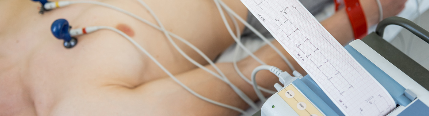
An electrophysiology study (EPS) is an invasive cardiac test in which thin catheters are passed through a vein or artery into the heart to record its electrical signals and deliberately stimulate it. The electrophysiology study helps identify the mechanism and location of an arrhythmia and guides treatment such as catheter ablation or device implantation.
주요 테이크 아웃
- An electrophysiology study maps the heart's electrical conduction, not its blood supply or pumping strength.
- An electrophysiology study is usually ordered after non-invasive tests such as ECG, Holter monitoring and echocardiography have been done.
- A negative or "non-inducible" electrophysiology study does not mean an arrhythmia can never occur.
- An electrophysiology study is often combined with catheter ablation in the same sitting, so consent usually covers both.
- Risks are uncommon but real, and differ between a diagnostic study and an ablation procedure. Results vary depending on individual clinical circumstances.
한눈에
| 제품 특장점 | 세부 |
|---|---|
| What is studied | Electrical conduction system and arrhythmia circuits of the heart |
| 접근 경로 | Usually femoral vein (groin); sometimes neck, subclavian or femoral artery |
| 마취 | Local anaesthetic at the puncture site, with sedation; general anaesthesia in selected cases |
| 일반적인 기간 | Diagnostic study commonly around 1-2 hours; longer, often 2-5 hours or more, if ablation is performed |
| 환경 | Cardiac electrophysiology (EP) lab or cath lab |
| 머무르다 | Day-care in some cases; overnight observation is common, especially after ablation or device implantation |
| 누가 수행합니까? | A cardiac electrophysiologist with EP lab staff |
라고도
- EP study, EPS
- Cardiac electrophysiology study
- Electrophysiological mapping / EP mapping
- Commonly described in India as the "heart electric test", "rhythm test" or "EP test", and often spoken of together with "ablation" or the "burning procedure"
- Related but distinct: "echo test" (echocardiography), "angiography" (coronary angiogram)
What an electrophysiology study is
The heartbeat is triggered by electrical impulses that begin in the sinus node, travel through the atria, pass through the AV node and His bundle, and spread through the ventricles. When this system misfires, conducts too slowly, or contains an extra pathway or a scar-related circuit, the result is an arrhythmia.
During an electrophysiology study, catheters with electrodes at their tips are positioned inside the heart chambers. These electrodes record local electrical signals from the inside, far more precisely than a surface ECG can. The electrophysiologist also paces the heart with tiny, timed electrical impulses (programmed stimulation) to measure conduction intervals and, where appropriate, to try to reproduce the patient's arrhythmia under controlled conditions so its mechanism can be identified.
Modern studies often use three-dimensional electroanatomical mapping systems, which build a computer model of the chamber and display where abnormal signals arise. If a treatable target is found, catheter ablation (radiofrequency energy, or cryoablation) may be delivered in the same procedure.
Why an electrophysiology study is done
- To define the mechanism of a documented arrhythmia: for example distinguishing types of supraventricular tachycardia (AVNRT, AV re-entrant tachycardia via an accessory pathway, atrial tachycardia) or localising ventricular tachycardia.
- To plan and guide catheter ablation, by pinpointing the circuit or focus to be targeted.
- To assess unexplained syncope when a rhythm cause is suspected but not captured on ECG or ambulatory monitoring, particularly with structural heart disease or conduction abnormalities.
- To evaluate conduction disease: for instance assessing AV or sinus node function when pacemaker need is uncertain.
- To help with risk assessment for ventricular arrhythmia in selected patients with scar-related heart disease or certain inherited channelopathies, as one input among many when considering an implantable cardioverter defibrillator (ICD).
- To assess an accessory pathway in Wolff-Parkinson-White pattern, including some athletes or people in safety-critical occupations.
- To reassess after a previous ablation when symptoms recur.
누가 이 검사를 받아야 할까요?
An electrophysiology study is a targeted, invasive investigation, it is not a screening or check-up test. It is generally considered for people who:
- have recurrent palpitations with a documented or strongly suspected tachyarrhythmia that needs mechanism-specific treatment
- are being considered for catheter ablation of SVT, atrial flutter, atrial fibrillation, ventricular tachycardia or frequent symptomatic ectopy
- have had syncope that is sudden, unexplained, exertional, or occurring with known structural or inherited heart disease
- have a WPW (accessory pathway) pattern on ECG with symptoms, or in some asymptomatic cases where occupational or sport risk matters
- have suspected but unproven conduction block causing symptoms
- are being assessed for ICD suitability in selected scar-related or inherited arrhythmia syndromes
일반적으로 지원 the right test for chest pain suspected to be angina, for breathlessness due to heart failure, or for palpitations that are clearly benign ectopy in a structurally normal heart, other tests answer those questions better. Not everyone with an arrhythmia needs an electrophysiology study; many are managed with medication and monitoring. Suitability is decided by the treating cardiologist or electrophysiologist.
What an electrophysiology study cannot detect or exclude
- 이 검사는 관상동맥을 평가하지 않습니다. An electrophysiology study does not show narrowing or blockage. A normal EPS does not exclude coronary artery disease; coronary angiography or CT coronary angiography answers that question.
- It does not measure pumping function or valves. Ejection fraction, valve disease and pericardial fluid are assessed by echocardiography or cardiac MRI, not by EPS.
- A non-inducible study does not exclude an arrhythmia. Some arrhythmias, notably many atrial fibrillation episodes, catecholamine-dependent rhythms and vagally mediated events, cannot be reliably provoked in the lab. Non-inducibility means no arrhythmia was triggered under the conditions tested; it is not the same as "you will never have one".
- It does not detect a cause outside the electrical system. Vasovagal or orthostatic faints, anaemia, thyroid disease, drug effects, anxiety-related palpitations and sleep apnoea are not diagnosed by EPS.
- It does not reliably predict future sudden cardiac death. Programmed stimulation has limited predictive value in several conditions, and prognosis depends on many other factors.
- It does not show tissue-level detail such as myocardial fibrosis, inflammation or infiltration; cardiac MRI is used for that.
If symptoms continue despite a normal or non-inducible electrophysiology study, return to your doctor. Further monitoring, extended Holter, patch monitor, or an implantable loop recorder, may be needed.
관련은 있지만 서로 다른 시험들
| Test | 질문에 대한 답변 | Key difference from EPS |
|---|---|---|
| 휴식 ECG | What is the rhythm and conduction right now? | Non-invasive, seconds-long snapshot; may miss intermittent arrhythmia |
| Holter / patch / extended ambulatory monitor | What rhythm occurs during daily life? | Non-invasive; captures spontaneous events but cannot define the circuit or treat it |
| 이식형 루프 레코더 | What rhythm occurs during rare, widely spaced events? | Long-term monitoring over months to years; diagnostic only |
| 운동(부하) 심전도 | Does exertion provoke ischaemia or arrhythmia? | Non-invasive; different physiology from lab pacing |
| Transthoracic echocardiography ("echo test") | Structure, valves, pumping function | Imaging of anatomy and function, not electrical circuits |
| 심장 MRI | Scar, fibrosis, inflammation, precise volumes | Tissue characterisation; no electrical mapping |
| 관상 동맥 조영술 | Are the coronary arteries narrowed? | Invasive but assesses blood supply, not conduction |
| Tilt-table test | Is fainting reflex/autonomic in origin? | Tests blood-pressure and heart-rate reflexes, not intracardiac signals |
| Diagnostic EPS vs catheter ablation | EPS diagnoses; ablation treats | Often done in one sitting, but they carry different risk profiles and consent needs |
These tests answer different questions. None is universally superior, the right first test depends on the presenting problem. For breathlessness with a murmur, echocardiography comes first. For infrequent palpitations, ambulatory monitoring comes first. For chest pain suspected to be ischaemic, coronary assessment comes first. An electrophysiology study is reserved for when the electrical mechanism itself must be defined or treated.
준비 방법
Preparation instructions are set by the performing unit and vary between hospitals and between operators. Your own unit's written instructions take precedence over anything on this page.
- Pre-procedure assessment: typically an examination, ECG, blood tests (including haemoglobin, renal function and clotting) and often an echocardiogram. Some units request imaging to exclude intracardiac thrombus before left-sided procedures.
- 금식: a period of fasting before sedation or anaesthesia is usual. The exact hours for solids and clear fluids are unit-specific.
- 의약품 : bring a complete list, including anti-arrhythmic drugs, blood thinners, diabetes medicines, inhalers, and any AYUSH, herbal or gym supplements. Some drugs are held before an electrophysiology study because they suppress inducibility. Do not start, stop or change any prescribed medicine yourself, only the prescriber or the EP team should advise this.
- Allergies and history: declare previous reactions to local anaesthetic, sedatives, antibiotics or contrast, plus bleeding tendencies, previous cardiac procedures and implanted devices.
- Skin and site preparation: the groin and sometimes chest or neck may be shaved and cleaned.
- 호송 및 운송: arrange for a responsible adult to accompany you home. Do not drive yourself on the day of sedation.
당뇨병이 있고 혈당 강하 치료를 받고 있다면
Fasting plus insulin or sulfonylureas can cause hypoglycaemia. Ask the team in advance exactly how to adjust timing and doses, and how blood glucose will be monitored on the day. Do not decide dose changes independently.
만약 만성 신장 질환이 있다면
Contrast may be used in some procedures. Tell the team your latest kidney function. Hydration protocols and contrast-sparing techniques are decided case by case, and fluid volumes must be individualised.
심부전이 있거나 수분 섭취가 제한된 경우
Generic "drink plenty of water" advice is not safe for everyone. Pre-procedure fluids and diuretic timing should be prescribed by your team, and lying flat for a long period may be uncomfortable, mention this beforehand.
If you are pregnant or could be
Electrophysiology studies use X-ray fluoroscopy. Tell the team before the procedure if pregnancy is possible. Where an arrhythmia must be treated during pregnancy, timing, shielding and low- or zero-fluoroscopy mapping techniques are considered; availability of such techniques varies by unit.
어린이와 청소년
Paediatric electrophysiology studies are performed in centres with paediatric electrophysiology expertise, usually under general anaesthesia, with catheter sizes, radiation doses and drug doses adjusted for the child. Fasting rules for children differ from adults.
고령자
Frailty, anticoagulation, kidney function, vascular disease and multiple medicines all influence planning and recovery. Sedation is often lighter and monitoring longer.
이식형 기기
Existing pacemakers or ICDs are interrogated before and after the procedure, and settings may be adjusted temporarily.
What happens during an electrophysiology study
- 설정: you lie on a narrow procedure table. ECG electrodes, a blood pressure cuff, oxygen saturation monitoring and an intravenous cannula are placed. Mapping patches may be applied to the chest and back.
- Local anaesthetic and access: the groin (or neck) is numbed. Using ultrasound guidance in many units, a needle enters the vein and sheaths are placed. Most people feel pressure rather than sharp pain.
- Catheter positioning: catheters are advanced to the heart under X-ray and/or 3D mapping guidance. Blood vessels have no pain nerves inside, so catheter movement is usually not painful.
- Recording and pacing: baseline intervals are measured, then programmed stimulation is delivered. You may feel your heart racing, thumping, fluttering, mild chest discomfort, lightheadedness or brief dizziness. Tell the team what you feel, it is useful information.
- Provoking and terminating arrhythmia: if an arrhythmia is induced, it is studied and then stopped by pacing, medication, or occasionally a brief synchronised shock while you are sedated.
- Drug challenge (selected cases): isoprenaline, adenosine or other agents may be given to unmask conduction or an arrhythmia.
- Ablation, if planned: once a target is confirmed, energy is applied. Brief discomfort in the chest, throat or shoulder can occur in some locations.
- 폐쇄: sheaths are removed and bleeding is controlled by manual pressure, a closure device or a suture. You will usually lie flat with the leg straight for a period defined by your unit.
과정이 얼마나 걸립니까
A purely diagnostic electrophysiology study commonly takes about one to two hours. When ablation is added, total lab time is often two to five hours, and complex cases such as ventricular tachycardia or persistent atrial fibrillation can take longer. Add preparation time before and several hours of monitored bed rest afterwards. Many patients go home the same day; overnight stay is common after ablation or device implantation. Actual duration and discharge timing depend on the findings and on unit protocol.
보고서 이해하기
An electrophysiology study report is a technical document written for the treating team. Typical elements include:
- 기준 측정: conduction intervals through the AV node and His-Purkinje system, sinus node recovery behaviour, and refractory periods. These are compared with accepted electrophysiological standards by the electrophysiologist; they are not values patients should interpret alone.
- Inducibility: whether an arrhythmia was provoked, what it was, how fast it was, how it started and stopped, and whether it reproduced your symptoms.
- Mechanism and location: for example AV nodal re-entry, an accessory pathway with its position, a focal atrial tachycardia, cavotricuspid isthmus-dependent flutter, or scar-related ventricular tachycardia.
- Ablation details: targets treated, energy used, and endpoints such as loss of pathway conduction, isthmus block, pulmonary vein isolation or non-inducibility.
- Conclusion and plan: for instance medication changes, repeat procedure, device implantation, or follow-up monitoring.
A "normal" study means no conduction abnormality was measured and no arrhythmia was induced on that day under those conditions. That is reassuring, but it does not by itself explain ongoing symptoms, and it does not exclude an arrhythmia that was simply not provoked. No result should be treated as a diagnosis on its own, it must be interpreted in your clinical context by the doctor who arranged the study.
결과가 오해를 불러일으킬 수 있는 경우
Falsely negative: arrhythmia present but not found
- Sedation and anaesthetic agents can suppress arrhythmia induction.
- Anti-arrhythmic drugs still in the system reduce inducibility.
- Arrhythmias that depend on adrenaline surges, exercise, sleep, hormonal cycles, alcohol or fever may not appear on a lab table.
- Autonomic or reflex causes of fainting are not detectable by intracardiac recording.
- Atrial fibrillation and some focal arrhythmias are poorly reproducible by programmed stimulation.
- Limited stimulation protocols, or an incompletely mapped chamber, can miss a circuit.
Falsely positive or clinically irrelevant findings
- Aggressive stimulation or high-dose isoprenaline can induce a rhythm that never occurs spontaneously, non-specific polymorphic ventricular arrhythmias being a classic example.
- A measured conduction abnormality may be an incidental finding rather than the cause of symptoms.
- An inducible arrhythmia that does not match your symptoms may not be the culprit.
- Catheter contact can cause mechanical ectopy or transient block that is mistaken for disease.
기타 오류 발생 원인
- Electrolyte disturbance, thyroid abnormality, anaemia, fever or recent alcohol can alter electrical behaviour on the day.
- Signal artefact, poor catheter contact or mapping-system registration error can distort a 3D map.
- Findings immediately after a fresh ablation can change as tissue oedema settles, so early non-inducibility does not guarantee permanent success.
위험 및 안전
Serious complications are uncommon in experienced hands, but an electrophysiology study is an invasive cardiac procedure and no such procedure is risk-free. Risk differs substantially between procedure types, and depends on your age, heart function, kidney function, anticoagulation and other illnesses. Results vary depending on individual clinical circumstances. Your operator should give you a personalised risk discussion before consent.
Diagnostic electrophysiology study
- Bruising, pain or haematoma at the groin or neck puncture site, the commonest issue
- Bleeding needing pressure, transfusion or, rarely, vascular repair; pseudoaneurysm or arteriovenous fistula
- Vein thrombosis; infection at the access site
- Induced arrhythmia requiring pacing, drugs or a brief shock to terminate
- Reaction to sedatives, local anaesthetic or drugs used for provocation
- Uncommon: cardiac perforation with pericardial effusion or tamponade; pneumothorax with neck or subclavian access
- Radiation exposure from fluoroscopy, minimised by modern low-dose and 3D mapping techniques
Right-sided ablation (for example AVNRT, atrial flutter, right-sided pathways)
- All of the above, plus a small risk of damage to the normal conduction system requiring a permanent pacemaker, the risk is higher when the target lies close to the AV node
- Localised chest discomfort or pericarditis-type pain after energy delivery
Left-sided ablation (for example atrial fibrillation, left-sided pathways, left ventricular VT)
- Requires arterial access or trans-septal puncture, adding risks of tamponade and vascular injury
- Stroke or transient ischaemic attack from clot or air embolism, reduced by anticoagulation and careful technique
- Procedure-specific risks such as phrenic nerve injury, pulmonary vein narrowing, and the rare but serious atrio-oesophageal fistula after atrial fibrillation ablation
- Higher likelihood of needing repeat procedures for persistent arrhythmia
Ventricular tachycardia ablation in structural heart disease
- Generally the highest-risk category, reflecting the underlying disease as much as the procedure
- Risks include haemodynamic deterioration during induced VT, need for mechanical support, kidney injury, longer procedures and greater radiation and contrast exposure
Red flags after an electrophysiology study
응급 상황: 즉시 진료를 받으십시오.
- Severe or worsening chest pain, pain radiating to the back, or pain with breathlessness
- 갑작스럽고 심한 호흡곤란 또는 안정 시 호흡곤란
- Fainting, near-fainting, or a sustained racing heartbeat with sweating or feeling about to collapse
- Sudden weakness or numbness of face, arm or leg, slurred speech, facial droop or sudden visual loss
- Heavy bleeding or a rapidly expanding, tense swelling at the puncture site, press firmly and get emergency help
- A cold, pale, numb or pulseless leg on the side of the puncture
- Repeated ICD shocks
Same day: contact your cardiac team today
- Fever above 38癈, or redness, spreading warmth, increasing tenderness or discharge at the access site
- A firm, painful, enlarging lump or a pulsating swelling in the groin
- New persistent palpitations, dizziness on standing, or recurrence of the original arrhythmia
- Persistent pain on swallowing, new heartburn or pain behind the breastbone after atrial fibrillation ablation
- Persistent hiccups, or breathlessness on lying flat, after ablation near the phrenic nerve
- Passing much less urine than usual after a procedure involving contrast
정기 진료
- Bruising that is spreading slowly but with no swelling, pain or fever
- Mild soreness or a small lump at the puncture site settling over days
- Occasional brief skipped beats or flutters, especially in the first weeks after ablation
- Questions about medicines, driving, exercise, or returning to work
특별한 상황
임신
Most arrhythmias in pregnancy are managed with medication and observation. Where an ablation is unavoidable, it is done in a specialised centre, ideally with minimised or zero fluoroscopy, and with obstetric input. Availability of low-radiation mapping varies between units.
어린이
Electrophysiology studies in children are usually performed under general anaesthesia by paediatric electrophysiologists. Accessory pathway tachycardias are the commonest indication. Growth, vessel size and cumulative radiation exposure all influence timing and technique, and some units prefer to defer elective procedures until the child is older.
고령자
Ablation can substantially reduce symptoms in older adults, but vascular access, bleeding risk, kidney function and cognitive status need careful assessment. Shorter, simpler procedures are often preferred.
혈당 강하 치료를 받는 당뇨병 환자
Prolonged fasting, sedation and delayed meals raise the risk of hypoglycaemia. Confirm insulin and tablet timing with your prescriber, and ensure glucose is checked before and after the procedure.
만성 신장 질환
Contrast and fluid load must be balanced. Anticoagulation dosing also changes with kidney function. Tell the team if you are on dialysis so timing can be coordinated.
심장 마비
Lying flat for a long period may be difficult, and fluid given during a long procedure can worsen congestion. Procedures may be staged, and diuretics adjusted by the treating team.
Existing pacemaker or ICD
Devices are checked before and after, and may be reprogrammed during the procedure. Bring your device identity card.
Electrophysiology study in the Indian context
- 운영시간: electrophysiology labs with 3D mapping are concentrated in larger cities and tertiary centres. Waiting times, the range of arrhythmias treated, and access to zero-fluoroscopy or cryoablation differ from centre to centre.
- 류마티스 성 심장병 remains an important cause of atrial arrhythmia in India. Atrial fibrillation and flutter in the setting of mitral valve disease behave differently from arrhythmia in a structurally normal heart, and treatment choices, including valve intervention and anticoagulation, are often addressed alongside rhythm management.
- Coronary disease at younger ages in South Asians means scar-related ventricular tachycardia can present earlier than expected; a previous heart attack should always be mentioned.
- 빈혈증, including iron-deficiency anaemia and thalassaemia trait, is common and can worsen palpitations. Correcting anaemia is part of assessment, not a substitute for it.
- Thyroid disease, high caffeine intake, tobacco, alcohol and gym or weight-loss supplements frequently contribute to palpitations and should be disclosed, since some stimulant supplements provoke arrhythmia.
- Operator volume matters. Complication rates for complex ablation are influenced by centre and operator experience; it is reasonable to ask how often your centre performs your specific procedure.
- 데이터 프라이버시 : hospital forms, patient portals and remote device monitoring involve personal health data, which is governed in India by the Digital Personal Data Protection Act, 2023. You may ask how your data is stored, shared and retained.
- 광고 및 주장: the Drugs and Magic Remedies (Objectionable Advertisements) Act, 1954 and NMC professional conduct rules prohibit guaranteed-cure claims. Treat any promise of a certain outcome with caution.
인도의 비용 및 보험
The cost of an electrophysiology study in India varies widely and cannot be quoted meaningfully as a single figure. A purely diagnostic study is the least expensive; adding ablation, 3D mapping, or a device implant increases cost substantially. Ask the hospital for a written, itemised estimate before admission, and confirm what is included.
비용에 영향을 미치는 요소:
- Diagnostic study alone versus study plus ablation, and whether one or several arrhythmias are treated
- Type of ablation, conventional radiofrequency, 3D-guided, cryoablation, or newer energy sources
- Number and type of catheters and sheaths, most of which are single-use consumables
- Use of a 3D electroanatomical mapping system, intracardiac echocardiography or trans-septal equipment
- Complexity: AVNRT or flutter versus persistent atrial fibrillation or scar-related ventricular tachycardia
- Anaesthesia type and duration, and length of lab time
- Room category and length of stay, including any intensive care
- Pre-procedure investigations and post-procedure follow-up, monitors or medicines
- Whether a pacemaker or ICD is implanted in the same admission
- City, hospital category, and whether the procedure is planned or emergency
- Repeat procedures, which are needed in a proportion of atrial fibrillation and VT cases
보험 확인 사항: most indemnity health insurance policies in India cover medically indicated cardiac electrophysiology procedures as inpatient treatment, but coverage is policy-specific. Confirm pre-authorisation requirements, waiting periods for pre-existing disease, room-rent capping (which can proportionately reduce the whole claim), consumable and implant sub-limits, and co-payment clauses. Government schemes such as Ayushman Bharat PM-JAY, CGHS, ECHS and state schemes have their own package rates and empanelment lists. Ask the hospital insurance desk to explain expected non-payable items in writing.
신화와 사실
| 신화 | 사실 |
|---|---|
| An electrophysiology study checks for heart blockages | An electrophysiology study examines electrical conduction. Coronary narrowing is assessed by coronary angiography or CT coronary angiography. |
| A normal EPS means you can never have a dangerous arrhythmia | A non-inducible study reflects that day's testing conditions. It lowers, but does not remove, the possibility of future arrhythmia. |
| Ablation permanently cures every arrhythmia in one sitting | Success rates differ greatly by arrhythmia type. Some, such as AVNRT, respond very well; persistent atrial fibrillation and scar-related VT often need repeat procedures or continued medication. Results vary depending on individual clinical circumstances. |
| Open-heart surgery is needed | An electrophysiology study is catheter-based through a blood vessel. The chest is not opened. |
| The procedure is done under full anaesthesia and you feel nothing at all | Many diagnostic studies use local anaesthetic with sedation. You may feel palpitations, pressure or brief dizziness, which is expected and monitored. |
| Feeling palpitations after ablation means the procedure failed | Brief skipped beats and short flutters are common in the early weeks while tissue heals. Persistent or sustained symptoms should be reported. |
| Every patient with palpitations needs an electrophysiology study | Most palpitations are investigated with ECG and ambulatory monitoring first. EPS is reserved for specific indications. |
| You must stop all heart medicines before the test | Only the prescriber or the EP team should change any medicine. Stopping drugs without advice can be dangerous. |
자주하는 질문
Is an electrophysiology study the same as an angiography?
No. An electrophysiology study records the heart's electrical signals through catheters placed inside the heart chambers. Coronary angiography injects contrast to show the coronary arteries. The two answer different questions, and one cannot substitute for the other, although both are catheter procedures done in a lab.
시술 중에 깨어있나요?
Often yes, in a drowsy, comfortable state after local anaesthetic and sedation. Staying partly awake can help, because your description of induced palpitations tells the team whether the arrhythmia matches your symptoms. General anaesthesia is used for children and for some complex ablations.
How soon can I return to normal activity?
Light activity usually resumes within a day or two after a diagnostic study. Heavy lifting, straining, driving and vigorous exercise are restricted for a period that depends on the access site, whether ablation was done, and your unit's advice. Follow your discharge instructions.
Is ablation always performed in the same sitting?
Not always. It is common to plan a combined study and ablation, and consent usually covers both. However, ablation may be deferred if no suitable target is found, if the arrhythm










 첸나이에서 가장 가까운 최고의 병원


