Geprüft vom Apollo-Redaktionsteam
Schnelle Antwort
3D heart mapping is a computer-guided technique that builds a three-dimensional picture of the inside of the heart and shows how electrical signals travel through it. Cardiologists use it during an electrophysiology (EP) study and catheter ablation to find the exact spot that is causing an abnormal heart rhythm. It is not a separate treatment or a scan you book on its own. It helps the doctor treat complex rhythm problems more precisely and with less X-ray exposure.
| Key facts | Details |
|---|---|
| Was es ist | A 3D electrical and anatomical map of a heart chamber, created with catheters and a computer system |
| Wann wird es verwendet? | During an EP study and catheter ablation for arrhythmia |
| Wer führt es durch | Ein Kardiologe mit Schwerpunkt Elektrophysiologie (Herzrhythmus-Kardiologe) |
| Zugriff | Thin catheters passed through a vein, and sometimes an artery, at the top of the leg |
| Anästhesie | Numbing injection with sedation, or general anaesthesia for longer procedures |
| Krankenhausaufenthalt | Same day or one night, depending on the ablation performed |
| Die wichtigsten Vorteile | Precise location of the rhythm problem; lower X-ray dose |
| Einschränkungen | Adds time and cost; depends on the abnormal rhythm being present or triggered during the procedure |
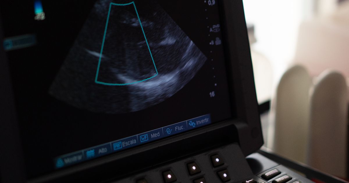
What is 3D heart mapping?
The heart beats because an electrical signal spreads through it in an orderly way. In an Arrhythmie, the signal starts in the wrong place, takes a short cut or circles around an area of scar. To treat the problem with ablation, the doctor first has to find exactly where this happens.
In conventional mapping, the doctor works this out from X-ray images and from electrical recordings taken by catheters placed at set positions in the heart. 3D electroanatomical mapping adds a tracking system. Patches on your chest and back, together with a weak magnetic field or tiny electrical currents, allow a computer to track the tip of a catheter in real time. As the catheter is moved along the inner wall of a chamber, it records its position and the local electrical signal at hundreds or thousands of points.
The computer joins these points into a three-dimensional shell of the chamber and colours it to show the timing or the strength of the electrical signal at each spot. The doctor can rotate this model, see where the abnormal rhythm begins or loops, and guide the ablation catheter to that place without continuous X-ray.
Other names for 3D heart mapping
- 3D mapping of the heart
- 3D electroanatomical mapping (EAM)
- Cardiac mapping or arrhythmia mapping
- 3D EP study
- 3D mapping-guided ablation
Why is 3D heart mapping done?
Mapping is the diagnostic part of an Elektrophysiologische Studie. Three-dimensional mapping is most useful when the rhythm problem is complex or lies in a part of the heart that is hard to picture on X-ray. It is commonly used for:
- Herzflimmern, particularly with radiofrequency ablation
- Atypisch Vorhofflattern und Vorhoftachykardie
- Ventrikuläre Tachykardie, especially when it arises from scar after a heart attack or in heart muscle disease
- Häufig vorzeitige ventrikuläre Kontraktionen that cause symptoms or weaken the heart
- Rhythm problems after heart surgery or in people born with a heart defect
- Repeat procedures after an earlier ablation
- Children, pregnant women and others in whom X-ray exposure should be kept as low as possible
Many straightforward rhythm problems, such as common forms of supraventrikuläre Tachykardie und Wolff-Parkinson-White-Syndrom, can be treated safely with conventional mapping. Not having 3D mapping does not mean you are receiving lesser care.
Types of maps and how mapping methods compare
The same system can display different kinds of information. The doctor chooses the maps that answer the question in your case.
| Kartentyp | Was es zeigt | Typische Verwendung |
|---|---|---|
| Anatomical map | The shape and size of the chamber and the vessels joining it | Guiding catheters and planning of ablation lines |
| Activation map | The order in which each area is electrically activated, shown as a colour sequence | Finding where a rhythm starts or the path of a circuit |
| Voltage map | The strength of the signal; low voltage suggests scar | Scar-related ventricular tachycardia and atrial scarring |
| Pace map | How closely a paced beat matches the abnormal beat on ECG | Extra beats that are too infrequent to map directly |
| Funktion | Conventional mapping | 3D-Zuordnung |
|---|---|---|
| Zielführung | X-ray and electrical recordings | Computer tracking plus electrical recordings, with X-ray as needed |
| Röntgenbelichtung | Höher | Normalerweise niedriger |
| Am besten geeignet, | Simple, well-defined rhythm problems | Complex, scar-related or repeat cases |
| Time and equipment | Weniger | More set-up, extra equipment and cost |
Images from a CT des Herzens or Herz-MRT taken beforehand can sometimes be merged with the map, and an ultrasound catheter inside the heart may be used alongside it.
Wer ist geeignet und wer möglicherweise nicht?
You may have 3D mapping if you are advised to undergo an EP study or Katheterablation and your cardiologist expects it to add useful information. The decision to use it is made by the operator, sometimes during the procedure itself.
The procedure may be postponed or avoided if:
- There is a blood clot inside the heart chamber to be mapped.
- You have an active infection or fever.
- You have a bleeding problem that has not been corrected.
- You are pregnant and the rhythm problem can safely wait; when it cannot, mapping helps to keep X-ray use very low.
- The rhythm problem is mild and well controlled with simpler treatment.
Wie man sich vorbereitet
Tests
- An EKG, ideally recorded during the abnormal rhythm.
- A Holter Monitor or Loop-Recorder to capture infrequent episodes.
- An Echokardiogramm um die Struktur und Funktion des Herzens zu beurteilen.
- Blood tests, and a CT or MRI scan in selected cases.
Zu besprechende Medikamente
Rhythm-control medicines are often paused for a few days beforehand so that the abnormal rhythm can be triggered and mapped. Blood thinners and diabetes medicines may also need a plan. Follow the written instructions from your team and do not stop anything on your own.
An dem Tag
- Do not eat for about six hours beforehand, or as instructed.
- Do not apply oil, lotion or powder to the chest and back, so that the mapping patches stick well.
- Leave jewellery at home and tell the staff about any implanted device or metal in your body.
- Organisieren Sie, dass Sie jemand nach Hause begleitet.
How is 3D heart mapping performed?
Mapping takes place in an electrophysiology laboratory. The length depends on the rhythm problem, from about an hour for a simple case to several hours for a complex one.
- Konfiguration: ECG stickers and the mapping patches are placed on your chest and back, and a drip is started.
- Anästhesie: the groin is numbed and sedation is given; some procedures are done under general anaesthesia.
- Katheterplatzierung: thin, flexible catheters are passed through the leg vein into the heart. For the left side of the heart, the doctor crosses the wall between the upper chambers or uses an artery.
- Building the shell: a mapping catheter is swept gently along the inner surface of the chamber while the computer draws its shape.
- Recording the signals: the abnormal rhythm is observed or triggered with small pacing impulses, and the timing and strength of signals are recorded across the chamber.
- Reading the map: the doctor studies the coloured map to find the source, the circuit or the scar channels responsible.
- Abtragung: if treatment is planned, energy is delivered at the target and each treated point is marked on the map. See Radiofrequenzablation des Herzens .
- Überprüfung: the team tries to trigger the rhythm again and may remap to confirm the result.
- Finishing: the catheters are removed and the puncture site is pressed or closed with a stitch.
Was passiert nach dem Eingriff?
You rest flat for a few hours while the puncture site seals, with your pulse, blood pressure and heart rhythm monitored. The mapping patches are removed. The doctor explains what the map showed, whether ablation was done and which medicines to take. Many people go home the same day or the next morning.
Risiken und Komplikationen
Mapping by itself adds very little risk. The risks are mainly those of the EP study and ablation it is part of, and they depend on the chamber treated.
Häufiger, in der Regel geringfügig
- Bruising or soreness at the groin
- Skin redness under the patches
- Palpitations or missed beats for a few days
Ungewöhnlich
- Bleeding or vessel injury at the groin
- Damage to the normal conduction system, which may require a pacemaker
- Blood collecting around the heart
- Stroke, mainly with procedures on the left side of the heart
- Blutgerinnsel in der Beinvene
- Reaction to sedation or anaesthesia
Einschränkungen, die man kennen sollte
- If the abnormal rhythm cannot be triggered on the day, the map may be incomplete.
- Movement and deep breathing can shift the map, so you may be asked to lie still.
- A detailed map helps the doctor but does not guarantee that ablation will work.
Ergebnisse: Wie gut funktioniert es?
3D mapping is a tool, so its value is judged by how it helps the ablation. Studies show that it reduces X-ray time and makes complex ablations more practical. For simple rhythm problems, success rates with conventional and 3D mapping are similar.
The outcome depends mainly on the rhythm being treated. Ablation works very well for many supraventricular tachycardias. Results are less certain for atrial fibrillation and scar-related ventricular tachycardia, where the rhythm may return and a repeat procedure or continued medicines may be needed. Some people with dangerous ventricular rhythms also need an implantierbarer Kardioverter-Defibrillator.
Erholung
- Erster Tag: rest; keep the groin dressing dry.
- Erste Woche: avoid heavy lifting, strenuous exercise and straining.
- Autofahren und Arbeit: many people return to light work within a few days; ask your doctor about driving.
- Nachverfolgen: a clinic visit with an ECG, and further monitoring if needed.
Wann sollte man medizinische Hilfe suchen?
- Bleeding or a rapidly enlarging swelling at the groin
- A cold, pale, numb or very painful leg
- Brustschmerzen oder Atemnot
- Ohnmacht oder starker Schwindel
- Fieber
- Weakness of the face, arm or leg, or difficulty speaking
- Schnelles Herzklopfen, das nicht nachlässt
Leben nach dem Eingriff
- Take medicines as prescribed, including blood thinners if advised.
- Keep a note of any palpitations, with the date, duration and what you were doing.
- Limit caffeine and alcohol if they trigger your symptoms.
- Manage blood pressure, weight, sleep and stress.
- Return to exercise gradually as your doctor advises.
Benefits of 3D heart mapping
- Shows the source or circuit of the rhythm problem in three dimensions
- Reduces X-ray exposure for you and the staff
- Identifies scar and helps plan ablation in damaged hearts
- Records where ablation has been applied, which helps avoid gaps
- Makes ablation possible for some complex rhythm problems
What is the cost of 3D heart mapping in India?
3D mapping is charged as part of the EP study and ablation. The cost depends on:
- The type and complexity of the rhythm problem
- The mapping patches and catheters used, most of which are single-use
- Whether ablation is performed and the energy source used
- Type of anaesthesia and procedure time
- Zimmerkategorie und Aufenthaltsdauer
- Scans done beforehand
- Die Stadt und das Krankenhaus
Die Kosten variieren je nach Person. Bitte erkundigen Sie sich im Krankenhaus nach den genauen Preisen.
Insurance cover for mapping equipment varies between policies, so check with your insurer before admission.
Why choose Apollo Hospitals for 3D heart mapping?
- Cardiologists trained in heart rhythm disorders who can advise whether mapping and ablation are needed
- Electrophysiology laboratories with anaesthesia and cardiac intensive care support
- Cardiac CT, MRI and echocardiography for planning
- Follow-up for rhythm monitoring, medicines and devices
- Die Verfügbarkeit variiert je nach Zentrum. Bitte erkundigen Sie sich im Krankenhaus.
To discuss your heart rhythm problem, Vereinbaren Sie einen Termin mit einem Kardiologen. in den Apollo-Krankenhäusern.
Verwandte lesen
- Elektrophysiologische Studie
- Katheterablation
- Katheterablation bei Vorhofflimmern
- Kryoballon-Ablation
- Gepulste Feldablation
Questions to ask your doctor about 3D heart mapping
- Do I need 3D mapping, or is conventional mapping enough for my rhythm problem?
- Will ablation be done in the same sitting?
- Which medicines should I stop beforehand, and when?
- Will I be sedated or under general anaesthesia?
- What is the chance of success for my type of arrhythmia?
- What happens if the rhythm cannot be triggered on the day?
- Was sind die Hauptrisiken in meinem Fall?
- How long will I stay in hospital, and when can I return to work?
Häufig gestellte Fragen
1. Is 3D heart mapping a treatment?
No. It is a guidance and diagnostic technique. The treatment is the ablation that usually follows in the same procedure.
2. Is 3D mapping the same as an EP study?
It is part of one. An EP study records the heart's electrical signals with catheters; 3D mapping displays those signals on a three-dimensional model of the chamber.
3. Is it like a CT or MRI scan?
No. CT and MRI are done from outside the body and show structure. 3D mapping is done with catheters inside the heart and shows electrical activity as well as shape.
4. Does everyone having ablation need 3D mapping?
No. Many simple rhythm problems are treated well with conventional mapping. 3D mapping is most helpful in complex, scar-related or repeat cases and when X-ray should be kept low.
5. Ist der Eingriff schmerzhaft?
The numbing injection stings briefly. After that, most people feel little, although you may notice palpitations when the rhythm is triggered and warmth or discomfort during ablation.
6. Wie lange dauert es?
From about an hour for a simple case to several hours for a complex one, including the ablation.
7. Does 3D mapping use radiation?
The mapping system itself does not. X-ray is still used for parts of the procedure, but usually much less than without 3D mapping.
8. Is it safe with a pacemaker or ICD?
Yes in most cases. The team takes care around the leads and checks the device afterwards.
9. Can children have 3D mapping?
Yes. It is often preferred in children because it lowers X-ray exposure. The procedure is usually done under general anaesthesia.
10. Will the map be accurate if I move?
Large movements can shift it. Sedation, a comfortable position and reference patches help, and the team can correct the map if needed.
11. What if my arrhythmia does not appear during the procedure?
The doctor may use pacing or medicines to bring it on. If it still cannot be triggered, mapping may rely on scar patterns or pace mapping, or the procedure may be repeated later.
12. Does 3D mapping improve the success of ablation?
It makes complex ablations more precise and practical. For simple rhythm problems, success is similar with or without it.
13. Wann kann ich zu meinen normalen Aktivitäten zurückkehren?
Most people resume light activities in a day or two and normal routines within about a week, depending on the ablation performed.
14. Which doctor performs 3D heart mapping?
A cardiac electrophysiologist, a cardiologist with additional training in heart rhythm disorders, performs it with a team of technicians, nurses and an anaesthetist.
Fazit
3D heart mapping gives the cardiologist a detailed, three-dimensional view of the heart's electrical activity during an EP study and ablation. It is especially useful for complex or scar-related rhythm problems and it lowers X-ray exposure, but it is a tool and not a treatment, and it is not needed in every case. Your heart rhythm specialist can tell you whether it is likely to help in your situation.
Diese Seite dient allgemeinen Informationen und ersetzt nicht die Beratung durch Ihren Arzt. Behandlungsentscheidungen sollten in Absprache mit einem qualifizierten Facharzt getroffen werden, der Ihre Krankengeschichte kennt.
Kürzlich Hinzugefügt


Herzspezialisten der Apollo-Kliniken







Haftungsausschluss:
Die Informationen auf dieser Seite dienen ausschließlich allgemeinen Informations- und Bildungszwecken. Obwohl wir angemessene Anstrengungen unternehmen, die Richtigkeit, Zuverlässigkeit und regelmäßige Überprüfung der Informationen zu gewährleisten, ersetzen diese keine professionelle medizinische Beratung, Diagnose oder Behandlung.
Die Eignung eines medizinischen Eingriffs sowie dessen Nutzen, Risiken, Vorbereitung, Genesung, mögliche Komplikationen und zu erwartende Ergebnisse können von Person zu Person variieren. Ihr Arzt wird anhand Ihres individuellen Zustands und Ihrer Krankengeschichte entscheiden, ob ein Eingriff für Sie geeignet ist.
Bitte konsultieren Sie vor jeder Entscheidung bezüglich eines medizinischen Eingriffs einen qualifizierten Arzt oder eine qualifizierte Ärztin, um eine individuelle Beratung zu erhalten.
Weitere Informationen darüber, wie unsere medizinischen Inhalte erstellt, geprüft, aktualisiert und gepflegt werden, finden Sie in unseren [Redaktionellen Richtlinien].