




Bild


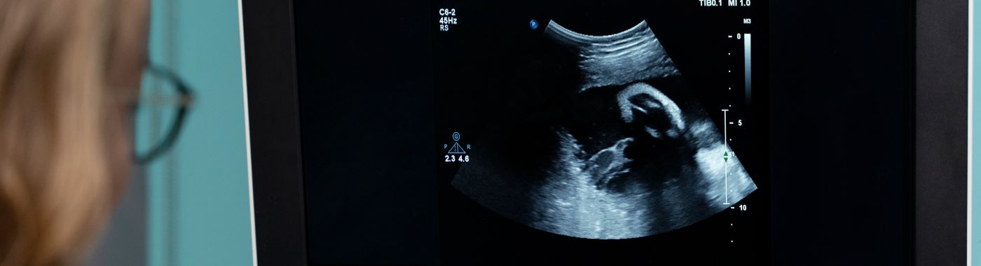
A 2D echo test, or two-dimensional echocardiography, is an ultrasound scan of the heart. The 2D echo shows heart chamber size, muscle thickness, pumping strength (ejection fraction), valve movement and fluid around the heart. A resting 2D echo does not image the coronary arteries, so a normal echo does not exclude coronary artery disease.
| Funktion | Detail |
|---|---|
| Testtyp | Non-invasive cardiac ultrasound (transthoracic echocardiography) |
| Strahlung | Keine Präsentation |
| Typical scan duration | About 15–45 minutes, longer if Doppler study is detailed, images are difficult or a contrast/stress protocol is added |
| Vorbereitung | Usually none for a standard resting echo; unit instructions take precedence |
| Needles or sedation | Not needed for a standard transthoracic 2D echo |
| Erholung | Normal activity immediately after a resting echo |
| Wer führt es durch | Cardiologist or trained cardiac sonographer; reported by a cardiologist |
| Hauptbeschränkung | Does not visualise coronary arteries; image quality is operator- and body-habitus dependent |
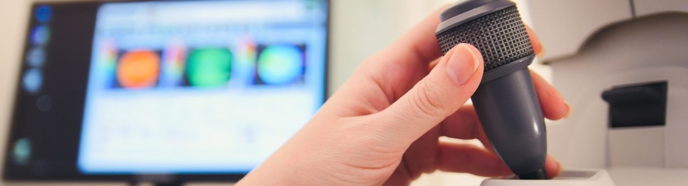
The 2D echo test uses high-frequency sound waves from a hand-held transducer placed on the chest. Sound waves reflect off heart muscle, valves and blood, and the machine converts the returning signals into moving two-dimensional images.
Most 2D echo studies in India are reported as 2D echo with colour Doppler. The 2D part shows anatomy and motion. Doppler measures the speed and direction of blood flow, which allows estimation of valve narrowing, valve leakage and some pressures inside the heart. M-mode tracings may also be included for chamber and wall measurements.
A 2D echo is a directed test, not a general health-check add-on. Consider discussing an echo with a doctor if you have:
An echo is not the right first test for everyone with chest pain, and a normal echo in a person with symptoms does not end the evaluation.
A finding not identified on an echo report means it was not seen on that study, not that it does not exist. If your symptoms persist or worsen despite a normal echo, go back to your doctor.
| Test | Fragen, die es beantwortet | Key difference from 2D echo |
|---|---|---|
| Resting 2D echo with colour Doppler | Is the heart's structure, valve function and pumping normal at rest? | Baseline anatomy and function; no coronary information |
| Stress echocardiography (exercise or dobutamine) | Does any heart wall stop moving well under stress, suggesting a flow-limiting coronary lesion? | Adds physiological stress; carries additional risk; needs monitoring and specific preparation |
| Transoesophageal echo (TEE/TOE) | Is there a clot in the left atrial appendage, endocarditis, prosthetic valve problem or subtle structural defect? | Probe passed into the oesophagus, usually with sedation and throat anaesthesia; invasive, requires fasting and consent |
| 3D-Echokardiographie | More detailed valve and chamber geometry, often before valve intervention | Supplementary to 2D, not a replacement |
| Contrast (agitated saline or ultrasound contrast) echo | Is there a shunt such as a patent foramen ovale, or is the left ventricle poorly visualised? | Requires an intravenous line and an agent; not part of a routine study |
| Fetale Echokardiographie | Does the unborn baby have a structural heart defect? | An obstetric ultrasound examination, legally distinct and governed by prenatal imaging regulations in India |
| EKG | What is the electrical rhythm and are there signs of ischaemia or chamber strain? | Electrical, not structural; takes minutes; no images |
| Coronary CT angiography or invasive coronary angiography | Are the coronary arteries narrowed, and where? | Directly images coronaries; involves radiation and iodinated contrast |
| Herz-MRT | Tissue characterisation, scar, infiltration, precise volumes | Longer, costlier, needs MRI safety assessment for implants |
There is no universally first cardiac test.
Preparation for a standard resting 2D echo is usually minimal. Protocols differ between hospitals and diagnostic centres, and the written instructions given by your own unit always take precedence over general advice.
Paediatric echo takes longer and may need a parent present, feeding or distraction. Sedation is occasionally used for uncooperative young children and is decided case by case.
A maternal 2D echo involves no radiation and is commonly performed during pregnancy when there is a cardiac concern. Fetal echocardiography is a separate examination performed by trained fetal-medicine or paediatric-cardiology specialists, and all prenatal ultrasound in India is governed by the PC-PNDT Act, 1994; sex determination is prohibited by law.
A resting echo needs no fasting, so meals and medicines continue normally. If your unit asks you to fast for a combined or stress study, ask the prescriber specifically how to handle insulin or sulfonylureas, as fasting on these drugs risks hypoglycaemia.
Lying flat may be uncomfortable if you are breathless; tell the sonographer, as most of the study can be done propped up or on your side. If you are on a fluid restriction, do not increase fluid intake for any scan instruction without checking with your treating doctor.
Turning on to the left side may be difficult with arthritis or frailty. Assistance and a slower pace are usual, and the examination can be modified.
Wie lange es dauert: a standard resting 2D echo with colour Doppler usually takes about 15–45 minutes, including positioning and measurements. Difficult imaging windows, detailed valve assessment, congenital heart disease and paediatric studies take longer. Transoesophageal and stress echo studies require additional preparation, monitoring and recovery time, so plan for a longer visit.
Echo reports are dense and technical. The sections below explain what the common headings refer to. No number on the report is a diagnosis by itself, and measurements must be read alongside your symptoms, examination and other tests by the doctor who requested the echo.
Ejection fraction estimates the proportion of blood ejected from the left ventricle with each beat. It is an estimate, not a precise measurement, and can vary between operators, between methods (visual estimate versus biplane volumes) and between visits. A borderline value in one report and a normal value in the next may reflect measurement variation rather than real change. Thresholds for normal, mildly reduced and reduced function should be read against the reference values quoted by your reporting laboratory and guideline edition in use.
Dilated chambers or thickened walls may point to long-standing high blood pressure, cardiomyopathy, valve disease or, in some cases, athletic adaptation. Measurements are influenced by body size, which matters particularly for South Asian patients, so indexed values are often used.
Valve narrowing (stenosis) and leakage (regurgitation) are usually graded as trivial, mild, moderate or severe. This grading is a composite judgement based on several Doppler measurements and images, and it is partly subjective and load-dependent — blood pressure, heart rate, anaemia and hydration all shift the numbers. Trivial or mild tricuspid or mitral regurgitation is an extremely common finding in healthy hearts and frequently needs no treatment at all. Patients often read any valve grade as serious disease; the grade must be interpreted with the clinical picture.
This describes how well the ventricle relaxes and fills. Reported patterns are indirect, influenced by age, rhythm and loading conditions, and are best treated as one piece of evidence rather than a standalone diagnosis.
Fluid around the heart may be described as minimal, mild, moderate or large. Significance depends on how quickly it collected and whether it is compressing the heart, not on volume alone.
Doppler-derived pulmonary artery pressure is an estimate, not a measured pressure, and depends on obtaining an adequate regurgitant jet. It can be over- or under-estimated.
Echo is operator-dependent. Two reports on the same heart, weeks apart, can differ slightly without any real change in the heart. Where a decision is borderline, your cardiologist may repeat the study, request a different echo type or use cardiac MRI.
A standard resting echo involves no radiation, no needles and no known tissue-damaging effects at diagnostic settings. The main discomforts are cold gel, probe pressure between the ribs, skin irritation from ECG stickers, and breathlessness if you must lie flat. It is widely used in newborns, pregnancy and older adults.
Stress echo carries genuine, though uncommon, risks because the heart is deliberately stressed: chest pain, arrhythmia, marked blood pressure change, and rarely serious cardiac events. It is performed with continuous monitoring, resuscitation facilities and specific consent. The safety profile of a resting echo does not apply to stress studies.
TEE is invasive. Risks include throat discomfort, sedation-related breathing and blood pressure effects, bleeding, and rarely oesophageal injury or perforation. Fasting and formal consent are required.
Ultrasound contrast agents are generally well tolerated but can cause hypersensitivity reactions and are used with caution in specific cardiac and pulmonary conditions. Agitated saline studies need an intravenous cannula.
Maternal echo is used when there is breathlessness beyond that expected in pregnancy, a murmur, known valve disease or previous cardiac surgery. Normal pregnancy itself increases blood volume and flow, which can slightly change chamber sizes and Doppler gradients; reports should be read with that in mind. Fetal heart assessment is a separate examination and is regulated under the PC-PNDT Act, 1994.
Paediatric echo is a distinct skill set focused on congenital anatomy. Image windows in infants are usually excellent. Findings such as a patent ductus in a newborn or a small septal defect need paediatric cardiology interpretation, as many close or remain harmless.
Age-related valve thickening, mild diastolic changes and mild aortic root dilatation are common and not automatically disease. Positioning limitations and lung changes may reduce image quality.
Beat-to-beat variation makes single measurements unreliable; averaging over several beats is required, and gradients may still be imprecise.
Fluid status strongly affects chamber sizes, pressures and valve gradients. Echoes done just before and just after dialysis can look quite different, so the timing should be recorded.
Serial echoes are used to track ventricular function and valve disease. Small changes in ejection fraction between studies may be measurement variation; treatment decisions are based on the trend plus symptoms.
Pacemakers, prosthetic valves and past bypass or valve surgery do not prevent an echo, but they alter images and require the reporting cardiologist to know the surgical history. If cardiac MRI is later considered, implants require an MRI safety assessment rather than being assumed unsafe.
A resting 2D echo with colour Doppler is one of the less expensive cardiac imaging tests, typically costing far less than CT coronary angiography or cardiac MRI. Prices differ widely between cities, between government and private facilities, and between standalone diagnostic centres and tertiary hospitals. Ask for the current charge at the point of booking; any figure quoted online is indicative only and may not reflect what you are charged.
Factors that change the price:
Versicherung: diagnostic tests done purely as outpatient investigations are often not payable under standard indemnity health policies unless the policy includes outpatient or diagnostic benefits. An echo performed during an admitted, covered hospitalisation is usually part of the claim. Coverage under government schemes and employer plans varies. Confirm with your insurer or the hospital's insurance desk before the test, and keep the prescription and report for any claim.
| Mythos | Tatsache |
|---|---|
| A normal 2D echo means my heart arteries are clear | A resting 2D echo does not image the coronary arteries. Assessing blockages needs stress testing, CT coronary angiography or invasive angiography. |
| An echo will show why my chest hurts | Echo answers structural questions. Many causes of chest pain — coronary, oesophageal, muscular, anxiety-related — are not visible on echo. |
| Echo uses X-rays, so it should be avoided in children and pregnancy | Echocardiography uses sound waves, not ionising radiation, and is routinely used in newborns and in pregnancy. |
| Mild valve leak on the report means heart disease | Trivial or mild mitral and tricuspid regurgitation is a frequent finding in healthy hearts and often needs no treatment. |
| A 3D echo is always better than 2D | 3D adds detail for specific valve and volume questions. It complements 2D rather than replacing it, and is not needed for most studies. |
| Ejection fraction is an exact number | Ejection fraction is an estimate that varies with method, operator and loading conditions. Trends matter more than single values. |
| An annual echo is a sensible routine screen for everyone | Echo is indicated when there is a clinical question. Screening healthy people without symptoms or risk indications mainly produces incidental findings and anxiety. |
A resting 2D echo is not painful for most people. The gel feels cold and the probe is pressed firmly between the ribs, which can be tender, particularly if your chest is sore or you are thin. Tell the sonographer if pressure becomes uncomfortable, as the angle can often be adjusted.
The scan itself usually takes about 15–45 minutes. Add time for registration, changing and waiting. Congenital, paediatric and technically difficult studies run longer, and stress or transoesophageal echo requires monitoring and recovery, which can extend the visit to a few hours.
No. The resting 2D echo does not show the coronary arteries and cannot predict a future heart attack. It may show muscle damage from a past attack. If coronary disease is suspected, your doctor will choose an ischaemia test or coronary imaging instead of relying on the echo.
Fasting is not usually needed for a resting echo. Fasting is commonly required for transoesophageal echo and some stress protocols. Follow the written instructions from your own unit, and if you take insulin or other glucose-lowering medicines, ask the prescriber how to manage them if fasting is requested.
No. An ECG records the heart's electrical activity in a few minutes and shows rhythm and some ischaemic or strain patterns. A 2D echo produces moving images of structure and pumping function. The two tests answer different questions and are frequently ordered together.
Report timing depends on the facility and whether a cardiologist reviews the images the same session. An abnormal finding is a starting point, not a verdict: your doctor will weigh it against your symptoms and examination and may repeat the echo, add other tests, or begin treatment.
Echo is most useful when there is a specific question — breathlessness, a murmur, known heart disease, abnormal ECG. In people without symptoms or indications, routine echo screening mostly yields incidental findings. Discuss with a doctor whether an echo adds anything in your situation.
Frequency depends on the condition and its severity. Mild, stable valve disease may be reviewed at long intervals; severe valve disease, cardiomyopathy or cancer treatment affecting the heart needs closer follow-up. Only your cardiologist can set the interval, based on your last study and current symptoms.
After a resting echo you can resume normal activity, driving and work straight away; there is no recovery period. After transoesophageal echo with sedation, you must not drive and will need an escort. Stress echo requires a period of observation before you leave.





 Bestes Krankenhaus in meiner Nähe in Chennai
Bestes Krankenhaus in meiner Nähe in Bangalore
Bestes Krankenhaus in meiner Nähe in Hyderabad
Bestes Krankenhaus in meiner Nähe in Mumbai
Bestes Krankenhaus in meiner Nähe in Delhi
Bestes Krankenhaus in meiner Nähe in Kolkata
Bestes Krankenhaus in meiner Nähe in Pune
Bestes Krankenhaus in meiner Nähe in Ahmedabad
Bestes Krankenhaus in meiner Nähe in Bhubaneswar
Bestes Krankenhaus in meiner Nähe in Bilaspur
Bestes Krankenhaus in meiner Nähe in Guwahati
Bestes Krankenhaus in meiner Nähe in Indore
Bestes Krankenhaus in meiner Nähe in Kochi
Bestes Krankenhaus in meiner Nähe in Lucknow
Bestes Krankenhaus in meiner Nähe in Madurai
Bestes Krankenhaus in meiner Nähe in Mysore
Bestes Krankenhaus in meiner Nähe in Nashik
Bestes Krankenhaus in meiner Nähe in Noida
Bestes Krankenhaus in meiner Nähe Rourkela
Bestes Krankenhaus in meiner Nähe in Visakhapatnam
Kardiologe in meiner Nähe in Chennai
Kardiologe in meiner Nähe in Bangalore
Kardiologe in meiner Nähe in Hyderabad
Kardiologe in meiner Nähe in Mumbai
Kardiologe in meiner Nähe in Delhi
Kardiologe in meiner Nähe in Kolkata
Kardiologe in meiner Nähe in Pune
Kardiologe in meiner Nähe in Ahmedabad
Kardiologe in meiner Nähe in Bhubaneswar
Kardiologe in meiner Nähe in Bilaspur
Kardiologe in meiner Nähe in Guwahati
Kardiologe in meiner Nähe in Indore
Kardiologe in meiner Nähe in Kochi
Kardiologe in meiner Nähe in Lucknow
Kardiologe in meiner Nähe in Madurai
Kardiologe in meiner Nähe in Mysore
Kardiologe in meiner Nähe in Nashik
Kardiologe in meiner Nähe in Noida
Kardiologe in meiner Nähe in Rourkela
Kardiologe in meiner Nähe in Visakhapatnam
Neurologe in meiner Nähe in Chennai
Neurologe in meiner Nähe in Bangalore
Neurologe in meiner Nähe in Hyderabad
Neurologe in meiner Nähe in Mumbai
Neurologe in meiner Nähe in Delhi
Neurologe in meiner Nähe in Kolkata
Neurologe in meiner Nähe in Ahmedabad
Neurologe in meiner Nähe in Bhubaneswar
Neurologe in meiner Nähe in Bilaspur
Neurologe in meiner Nähe in Guwahati
Neurologe in meiner Nähe in Indore
Neurologe in meiner Nähe in Kochi
Neurologe in meiner Nähe in Lucknow
Neurologe in meiner Nähe in Madurai
Neurologe in meiner Nähe in Mysore
Neurologe in meiner Nähe in Nashik
Neurologe in meiner Nähe in Noida
Neurologe in meiner Nähe in Rourkela
Neurologe in meiner Nähe in Visakhapatnam
Orthopäde in meiner Nähe in Chennai
Orthopäde in meiner Nähe in Bangalore
Orthopäde in meiner Nähe in Hyderabad
Orthopäde in meiner Nähe in Mumbai
Orthopäde in meiner Nähe in Delhi
Orthopäde in meiner Nähe in Kolkata
Orthopäde in meiner Nähe in Pune
Orthopäde in meiner Nähe in Ahmedabad
Orthopäde in meiner Nähe in Bhubaneswar
Orthopäde in meiner Nähe in Bilaspur
Orthopäde in meiner Nähe in Guwahati
Orthopäde in meiner Nähe in Indore
Orthopäde in meiner Nähe in Kochi
Orthopäde in meiner Nähe in Lucknow
Orthopäde in meiner Nähe in Madurai
Orthopäde in meiner Nähe in Mysore
Orthopäde in meiner Nähe in Nashik
Orthopäde in meiner Nähe in Noida
Orthopäde in meiner Nähe in Rourkela
Orthopäde in meiner Nähe in Visakhapatnam
Onkologe in meiner Nähe in Chennai
Onkologe in meiner Nähe in Bangalore
Onkologe in meiner Nähe in Hyderabad
Onkologe in meiner Nähe in Mumbai
Onkologe in meiner Nähe in Delhi
Onkologe in meiner Nähe in Kolkata
Onkologe in meiner Nähe in Pune
Onkologe in meiner Nähe in Ahmedabad
Onkologe in meiner Nähe in Bhubaneswar
Onkologe in meiner Nähe in Bilaspur
Onkologe in meiner Nähe in Guwahati
Onkologe in meiner Nähe in Indore
Onkologe in meiner Nähe in Kochi
Onkologe in meiner Nähe in Lucknow
Onkologe in meiner Nähe in Madurai
Onkologe in meiner Nähe in Mysore
Onkologe in meiner Nähe in Nashik
Onkologe in meiner Nähe in Noida
Onkologe in meiner Nähe in Rourkela
Gastroenterologe in meiner Nähe in Chennai
Gastroenterologe in meiner Nähe in Bangalore
Gastroenterologe in meiner Nähe in Hyderabad
Gastroenterologe in meiner Nähe in Mumbai
Gastroenterologe in meiner Nähe in Delhi
Gastroenterologe in meiner Nähe in Kolkata
Gastroenterologe in meiner Nähe in Pune
Gastroenterologe in meiner Nähe in Ahmedabad
Gastroenterologe in meiner Nähe in Bhubaneswar
Gastroenterologe in meiner Nähe in Bilaspur
Gastroenterologe in meiner Nähe in Guwahati
Gastroenterologe in meiner Nähe in Indore
Gastroenterologe in meiner Nähe in Kochi
Gastroenterologe in meiner Nähe in Lucknow
Gastroenterologe in meiner Nähe in Madurai
Gastroenterologe in meiner Nähe in Mysore
Gastroenterologe in meiner Nähe in Nashik
Gastroenterologe in meiner Nähe in Noida
Gastroenterologe in meiner Nähe in Rourkela
Gastroenterologe in meiner Nähe in Visakhapatnam
Transplantationschirurg in meiner Nähe in Chennai
Transplantationschirurg in meiner Nähe in Bangalore
Transplantationschirurg in meiner Nähe in Hyderabad
Transplantationschirurg in meiner Nähe in Mumbai
Transplantationschirurg in meiner Nähe in Delhi
Transplantationschirurg in meiner Nähe in Kolkata
Transplantationschirurg in meiner Nähe in Indore
Transplantationschirurg in meiner Nähe in Kochi
Transplantationschirurg in meiner Nähe in Lucknow
Transplantationschirurg in meiner Nähe in Mysore
HNO-Arzt in meiner Nähe in Chennai
HNO-Arzt in meiner Nähe in Bangalore
HNO-Arzt in meiner Nähe in Hyderabad
HNO-Arzt in meiner Nähe in Mumbai
HNO-Arzt in meiner Nähe in Delhi
HNO-Arzt in meiner Nähe in Kolkata
HNO-Arzt in meiner Nähe in Pune
HNO-Arzt in meiner Nähe in Ahmedabad
HNO-Arzt in meiner Nähe in Bhubaneswar
HNO-Arzt in meiner Nähe in Bilaspur
HNO-Arzt in meiner Nähe in Guwahati
HNO-Arzt in meiner Nähe in Indore
HNO-Arzt in meiner Nähe in Kochi
HNO-Arzt in meiner Nähe in Lucknow
HNO-Arzt in meiner Nähe in Madurai
HNO-Arzt in meiner Nähe in Mysore
HNO-Arzt in meiner Nähe in Nashik
HNO-Arzt in meiner Nähe in Noida
HNO-Arzt in meiner Nähe in Rourkela
HNO-Arzt in meiner Nähe in Visakhapatnam
Lungenfacharzt in meiner Nähe in Chennai
Lungenfacharzt in meiner Nähe in Bangalore
Lungenfacharzt in meiner Nähe in Hyderabad
Lungenfacharzt in meiner Nähe in Mumbai
Lungenfacharzt in meiner Nähe in Delhi
Lungenfacharzt in meiner Nähe in Kolkata
Lungenfacharzt in meiner Nähe in Ahmedabad
Lungenfacharzt in meiner Nähe in Bhubaneswar
Lungenfacharzt in meiner Nähe in Bilaspur
Lungenfacharzt in meiner Nähe in Guwahati
Lungenfacharzt in meiner Nähe in Indore
Lungenfacharzt in meiner Nähe in Kochi
Lungenfacharzt in meiner Nähe in Lucknow
Lungenfacharzt in meiner Nähe in Madurai
Lungenfacharzt in meiner Nähe in Mysore
Lungenfacharzt in meiner Nähe in Nashik
Lungenfacharzt in meiner Nähe in Noida
Lungenarzt in meiner Nähe in Rourkela
Lungenfacharzt in meiner Nähe in Visakhapatnam
Zahnarzt in meiner Nähe in Chennai
Zahnarzt in meiner Nähe in Bangalore
Zahnarzt in meiner Nähe in Hyderabad
Zahnarzt in meiner Nähe in Delhi
Zahnarzt in meiner Nähe in Kolkata
Zahnarzt in meiner Nähe in Ahmedabad
Zahnarzt in meiner Nähe in Bhubaneswar
Zahnarzt in meiner Nähe in Bilaspur
Zahnarzt in meiner Nähe in Guwahati
Zahnarzt in meiner Nähe in Kochi
Zahnarzt in meiner Nähe in Lucknow
Zahnarzt in meiner Nähe in Madurai
Zahnarzt in meiner Nähe in Nashik
Zahnarzt in meiner Nähe in Noida
Zahnarzt in meiner Nähe in Rourkela
Zahnarzt in meiner Nähe in Visakhapatnam
Kinderarzt in meiner Nähe in Chennai
Kinderarzt in meiner Nähe in Bangalore
Kinderarzt in meiner Nähe in Hyderabad
Kinderarzt in meiner Nähe in Mumbai
Kinderarzt in meiner Nähe in Delhi
Kinderarzt in meiner Nähe in Kolkata
Kinderarzt in meiner Nähe in Pune
Kinderarzt in meiner Nähe in Ahmedabad
Kinderarzt in meiner Nähe in Bhubaneswar
Kinderarzt in meiner Nähe in Bilaspur
Kinderarzt in meiner Nähe in Guwahati
Kinderarzt in meiner Nähe in Indore
Kinderarzt in meiner Nähe in Kochi
Kinderarzt in meiner Nähe in Lucknow
Kinderarzt in meiner Nähe in Madurai
Kinderarzt in meiner Nähe in Mysore
Kinderarzt in meiner Nähe in Nashik
Kinderarzt in meiner Nähe in Noida
Kinderarzt in meiner Nähe in Visakhapatnam
Dermatologe in meiner Nähe in Chennai
Dermatologe in meiner Nähe in Bangalore
Dermatologe in meiner Nähe in Hyderabad
Dermatologe in meiner Nähe in Delhi
Dermatologe in meiner Nähe in Kolkata
Dermatologe in meiner Nähe in Ahmedabad
Dermatologe in meiner Nähe in Bhubaneswar
Dermatologe in meiner Nähe in Bilaspur
Dermatologe in meiner Nähe in Guwahati
Dermatologe in meiner Nähe in Indore
Dermatologe in meiner Nähe in Kochi
Dermatologe in meiner Nähe in Lucknow
Dermatologe in meiner Nähe in Madurai
Dermatologe in meiner Nähe in Mysore
Dermatologe in meiner Nähe in Noida
Dermatologe in meiner Nähe in Visakhapatnam
Urologe in meiner Nähe in Chennai
Urologe in meiner Nähe in Bangalore
Urologe in meiner Nähe in Hyderabad
Urologe in meiner Nähe in Delhi
Urologe in meiner Nähe in Kolkata
Urologe in meiner Nähe in Pune
Urologe in meiner Nähe in Ahmedabad
Urologe in meiner Nähe in Bhubaneswar
Urologe in meiner Nähe in Bilaspur
Urologe in meiner Nähe in Guwahati
Urologe in meiner Nähe in Indore
Urologe in meiner Nähe in Kochi
Urologe in meiner Nähe in Lucknow
Urologe in meiner Nähe in Madurai
Urologe in meiner Nähe in Mysore
Urologe in meiner Nähe in Nashik
Urologe in meiner Nähe in Noida
Urologe in meiner Nähe in Rourkela
Urologe in meiner Nähe in Visakhapatnam
Endokrinologe und Diabetologe in meiner Nähe in Chennai
Endokrinologe und Diabetologe in meiner Nähe in Bangalore
Endokrinologe und Diabetologe in meiner Nähe in Hyderabad
Endokrinologe und Diabetologe in meiner Nähe in Mumbai
Endokrinologe und Diabetologe in meiner Nähe in Delhi
Endokrinologe und Diabetologe in meiner Nähe in Kolkata
Endokrinologe und Diabetologe in meiner Nähe in Pune
Endokrinologe und Diabetologe in meiner Nähe in Ahmedabad
Endokrinologe und Diabetologe in meiner Nähe in Bhubaneswar
Endokrinologe und Diabetologe in meiner Nähe in Bilaspur
Endokrinologe und Diabetologe in meiner Nähe in Guwahati
Endokrinologe und Diabetologe in meiner Nähe in Indore
Endokrinologe und Diabetologe in meiner Nähe in Kochi
Endokrinologe und Diabetologe in meiner Nähe in Lucknow
Endokrinologe und Diabetologe in meiner Nähe in Madurai
Endokrinologe und Diabetologe in meiner Nähe in Mysore
Endokrinologe und Diabetologe in meiner Nähe in Noida
Endokrinologe und Diabetologe in meiner Nähe in Rourkela
Endokrinologe und Diabetologe in meiner Nähe in Visakhapatnam
Frauenarzt in meiner Nähe in Chennai
Frauenarzt in meiner Nähe in Bangalore
Frauenarzt in meiner Nähe in Hyderabad
Frauenarzt in meiner Nähe in Mumbai
Frauenarzt in meiner Nähe in Delhi
Frauenarzt in meiner Nähe in Kolkata
Frauenarzt in meiner Nähe in Ahmedabad
Frauenarzt in meiner Nähe in Bhubaneswar
Frauenarzt in meiner Nähe in Bilaspur
Frauenarzt in meiner Nähe in Guwahati
Frauenarzt in meiner Nähe in Indore
Frauenarzt in meiner Nähe in Kochi
Frauenarzt in meiner Nähe in Lucknow
Frauenarzt in meiner Nähe in Madurai
Frauenarzt in meiner Nähe in Mysore
Frauenarzt in meiner Nähe in Nashik
Frauenarzt in meiner Nähe in Noida
Frauenarzt in meiner Nähe in Rourkela
Frauenarzt in meiner Nähe in Visakhapatnam
Allgemeinmediziner in meiner Nähe in Chennai
Allgemeinmediziner in meiner Nähe in Bangalore
Allgemeinmediziner in meiner Nähe in Hyderabad
Allgemeinmediziner in meiner Nähe in Mumbai
Allgemeinmediziner in meiner Nähe in Delhi
Allgemeinmediziner in meiner Nähe in Kolkata
Allgemeinmediziner in meiner Nähe in Pune
Allgemeinmediziner in meiner Nähe in Ahmedabad
Allgemeinmediziner in meiner Nähe in Bhubaneswar
Allgemeinmediziner in meiner Nähe in Bilaspur
Allgemeinmediziner in meiner Nähe in Guwahati
Allgemeinmediziner in meiner Nähe in Indore
Allgemeinmediziner in meiner Nähe in Kochi
Allgemeinmediziner in meiner Nähe in Lucknow
Allgemeinmediziner in meiner Nähe in Madurai
Allgemeinmediziner in meiner Nähe in Mysore
Allgemeinmediziner in meiner Nähe in Nashik
Allgemeinmediziner in meiner Nähe in Noida
Allgemeinmediziner in meiner Nähe in Visakhapatnam
Psychologe in meiner Nähe in Chennai
Psychologe in meiner Nähe in Bangalore
Psychologe in meiner Nähe in Hyderabad
Psychologe in meiner Nähe in Mumbai
Psychologe in meiner Nähe in Delhi
Psychologe in meiner Nähe in Kolkata
Psychologe in meiner Nähe in Ahmedabad
Psychologe in meiner Nähe in Bhubaneswar
Psychologe in meiner Nähe in Bilaspur
Psychologe in meiner Nähe in Guwahati
Psychologe in meiner Nähe in Indore
Psychologe in meiner Nähe in Kochi
Psychologe in meiner Nähe in Lucknow
Psychologe in meiner Nähe in Madurai
Psychologe in meiner Nähe in Mysore
Psychologe in meiner Nähe in Nashik
Psychologe in meiner Nähe in Noida
Psychologe in meiner Nähe in Visakhapatnam
Allgemeinchirurg in meiner Nähe in Chennai
Allgemeinchirurg in meiner Nähe in Bangalore
Allgemeinchirurg in meiner Nähe in Hyderabad
Allgemeinchirurg in meiner Nähe in Mumbai
Allgemeinchirurg in meiner Nähe in Delhi
Allgemeinchirurg in meiner Nähe in Kolkata
Allgemeinchirurg in meiner Nähe in Pune
Allgemeinchirurg in meiner Nähe in Ahmedabad
Allgemeinchirurg in meiner Nähe in Bhubaneswar
Allgemeinchirurg in meiner Nähe in Bilaspur
Allgemeinchirurg in meiner Nähe in Guwahati
Allgemeinchirurg in meiner Nähe in Indore
Allgemeinchirurg in meiner Nähe in Lucknow
Allgemeinchirurg in meiner Nähe in Madurai
Allgemeinchirurg in meiner Nähe in Mysore
Allgemeinchirurg in meiner Nähe in Nashik
Allgemeinchirurg in meiner Nähe in Noida
Allgemeinchirurg in meiner Nähe in Rourkela
Bestes Krankenhaus in Karukutty, Cochin
Bestes Krankenhaus in der Greams Road, Chennai
Bestes Krankenhaus in Kuvempunagar, Mysore
Bestes Krankenhaus in Vanagaram, Chennai
Bestes Krankenhaus in Teynampet, Chennai
Bestes Krankenhaus in OMR, Chennai
Bestes Krebskrankenhaus in Bhat, Gandhinagar, Ahmedabad
Bestes Krebskrankenhaus in Electronic City, Bangalore
Bestes Krebskrankenhaus in Teynampet, Chennai
Bestes Krebskrankenhaus in HSR Layout, Bangalore
Bestes Protonen-Krebszentrum in Chennai
Bestes Kinderkrankenhaus in Thousand Lights, Chennai
Bestes Frauenkrankenhaus in Thousand Lights, Chennai
Bestes Krankenhaus in Paschim Boragaon, Guwahati
Bestes Krankenhaus in der PH Road, Chennai
Bestes Herzzentrum in Thousand Lights, Chennai
Bestes Krankenhaus in Jubilee Hills, Hyderabad
Bestes Krankenhaus in Tondiarpet, Chennai
Bestes Krankenhaus in Kotturpuram, Chennai
Bestes Krankenhaus in der Kovai Road, Karur
Bestes Krankenhaus in Karapakkam, Chennai
Bestes Krankenhaus in Arilova, Vizag
Bestes Krankenhaus an der Kanpur Road, Lucknow
Bestes Krankenhaus in Sektor 26, Noida
Bestes Krankenhaus in Gandhinagar, Ahmedabad
Bestes Krankenhaus in Aragonda, Andhra Pradesh
Bestes Krankenhaus an der Bannerghatta Road, Bangalore
Bestes Krankenhaus in Unit-15, Bhubaneswar
Bestes Krankenhaus an der Seepat Road, Bilaspur
Bestes Krankenhaus in Ellisbridge, Ahmedabad
Bestes Krankenhaus in Neu-Delhi
Bestes Krankenhaus in DRDO, Hyderabad
Bestes Krankenhaus in der GS Road, Guwahati
Bestes Krankenhaus in Hyderguda, Hyderabad
Bestes Krankenhaus in Vijay Nagar, Indore
Bestes Krankenhaus an der Suryaraopeta Main Road, Kakinada
Bestes Krankenhaus an der Canal Circular Road, Kolkata
Bestes Krankenhaus im CBD Belapur, Navi Mumbai
Bestes Krankenhaus in Panchavati, Nashik
Bestes Krankenhaus in Secunderabad, Hyderabad
Bestes Krankenhaus in Seshadripuram, Bangalore
Bestes Krankenhaus an der Waltair Main Road, Visakhapatnam
Bestes Krankenhaus in der Subhash Nagar Road, Karimnagar
Bestes Krankenhaus in Managari, Karaikudi
Bestes Krankenhaus in Arepally, Warangal
Bestes Krankenhaus in Arera Colony, Bhopal
Bestes Krankenhaus in Jayanagar, Bangalore
Bestes Krankenhaus in KK Nagar, Madurai
Bestes Krankenhaus in Ramji Nagar, Nellore
Bestes Krankenhaus im Sektor 19, Rourkela
Bestes Krankenhaus in Swargate, Pune
Bestes Frauenkrebskrankenhaus in Süd-Delhi
CABG
CAR T-Zelltherapie
Laparoskopische Cholezystektomie
Hysterektomie
Nierentransplantation
Extrakorporale Stoßwellenlithotripsie
Leber-Transplantation
Lungentransplantation
Hüftarthroskopie
Vollständige Hüfterneuerung
Protonentherapie
Minimalinvasiver Subvastus-Kniegelenkersatz
Knieersatz in der Kindertagesstätte im Schnelldurchgang
Sleeve-Gastrektomie
Lasik-Operation
Nasenkorrektur
Fettabsaugung
Koronarangiogramm
Transkatheter-Aortenklappenersatz
MitraClip-Ventilreparatur
Minimal Invasive Herzchirurgie
Katheterablation
ACL-Rekonstruktionsoperation
Reverse-Shoulder-Ersatz
Endometriumablation
Uterusarterienembolisation
Ovarialzystektomie
Brustkrebs-Chirurgie
Brachytherapie
Koloskopie
Polypektomie
Tiefenhirnstimulation
Peritonealdialyse
Nierenbiopsie
Parathyreoidektomie
Zytoreduktive Chirurgie
Keramik-Totalersatz des Knies
ERCP
Bestes Krankenhaus in meiner Nähe in Chennai
Bestes Krankenhaus in meiner Nähe in Bangalore
Bestes Krankenhaus in meiner Nähe in Hyderabad
Bestes Krankenhaus in meiner Nähe in Mumbai
Bestes Krankenhaus in meiner Nähe in Delhi
Bestes Krankenhaus in meiner Nähe in Kolkata
Bestes Krankenhaus in meiner Nähe in Pune
Bestes Krankenhaus in meiner Nähe in Ahmedabad
Bestes Krankenhaus in meiner Nähe in Bhubaneswar
Bestes Krankenhaus in meiner Nähe in Bilaspur
Bestes Krankenhaus in meiner Nähe in Guwahati
Bestes Krankenhaus in meiner Nähe in Indore
Bestes Krankenhaus in meiner Nähe in Kochi
Bestes Krankenhaus in meiner Nähe in Lucknow
Bestes Krankenhaus in meiner Nähe in Madurai
Bestes Krankenhaus in meiner Nähe in Mysore
Bestes Krankenhaus in meiner Nähe in Nashik
Bestes Krankenhaus in meiner Nähe in Noida
Bestes Krankenhaus in meiner Nähe Rourkela
Bestes Krankenhaus in meiner Nähe in Visakhapatnam
Kardiologe in meiner Nähe in Chennai
Kardiologe in meiner Nähe in Bangalore
Kardiologe in meiner Nähe in Hyderabad
Kardiologe in meiner Nähe in Mumbai
Kardiologe in meiner Nähe in Delhi
Kardiologe in meiner Nähe in Kolkata
Kardiologe in meiner Nähe in Pune
Kardiologe in meiner Nähe in Ahmedabad
Kardiologe in meiner Nähe in Bhubaneswar
Kardiologe in meiner Nähe in Bilaspur
Kardiologe in meiner Nähe in Guwahati
Kardiologe in meiner Nähe in Indore
Kardiologe in meiner Nähe in Kochi
Kardiologe in meiner Nähe in Lucknow
Kardiologe in meiner Nähe in Madurai
Kardiologe in meiner Nähe in Mysore
Kardiologe in meiner Nähe in Nashik
Kardiologe in meiner Nähe in Noida
Kardiologe in meiner Nähe in Rourkela
Kardiologe in meiner Nähe in Visakhapatnam
Neurologe in meiner Nähe in Chennai
Neurologe in meiner Nähe in Bangalore
Neurologe in meiner Nähe in Hyderabad
Neurologe in meiner Nähe in Mumbai
Neurologe in meiner Nähe in Delhi
Neurologe in meiner Nähe in Kolkata
Neurologe in meiner Nähe in Ahmedabad
Neurologe in meiner Nähe in Bhubaneswar
Neurologe in meiner Nähe in Bilaspur
Neurologe in meiner Nähe in Guwahati
Neurologe in meiner Nähe in Indore
Neurologe in meiner Nähe in Kochi
Neurologe in meiner Nähe in Lucknow
Neurologe in meiner Nähe in Madurai
Neurologe in meiner Nähe in Mysore
Neurologe in meiner Nähe in Nashik
Neurologe in meiner Nähe in Noida
Neurologe in meiner Nähe in Rourkela
Neurologe in meiner Nähe in Visakhapatnam
Orthopäde in meiner Nähe in Chennai
Orthopäde in meiner Nähe in Bangalore
Orthopäde in meiner Nähe in Hyderabad
Orthopäde in meiner Nähe in Mumbai
Orthopäde in meiner Nähe in Delhi
Orthopäde in meiner Nähe in Kolkata
Orthopäde in meiner Nähe in Pune
Orthopäde in meiner Nähe in Ahmedabad
Orthopäde in meiner Nähe in Bhubaneswar
Orthopäde in meiner Nähe in Bilaspur
Orthopäde in meiner Nähe in Guwahati
Orthopäde in meiner Nähe in Indore
Orthopäde in meiner Nähe in Kochi
Orthopäde in meiner Nähe in Lucknow
Orthopäde in meiner Nähe in Madurai
Orthopäde in meiner Nähe in Mysore
Orthopäde in meiner Nähe in Nashik
Orthopäde in meiner Nähe in Noida
Orthopäde in meiner Nähe in Rourkela
Orthopäde in meiner Nähe in Visakhapatnam
Onkologe in meiner Nähe in Chennai
Onkologe in meiner Nähe in Bangalore
Onkologe in meiner Nähe in Hyderabad
Onkologe in meiner Nähe in Mumbai
Onkologe in meiner Nähe in Delhi
Onkologe in meiner Nähe in Kolkata
Onkologe in meiner Nähe in Pune
Onkologe in meiner Nähe in Ahmedabad
Onkologe in meiner Nähe in Bhubaneswar
Onkologe in meiner Nähe in Bilaspur
Onkologe in meiner Nähe in Guwahati
Onkologe in meiner Nähe in Indore
Onkologe in meiner Nähe in Kochi
Onkologe in meiner Nähe in Lucknow
Onkologe in meiner Nähe in Madurai
Onkologe in meiner Nähe in Mysore
Onkologe in meiner Nähe in Nashik
Onkologe in meiner Nähe in Noida
Onkologe in meiner Nähe in Rourkela
Gastroenterologe in meiner Nähe in Chennai
Gastroenterologe in meiner Nähe in Bangalore
Gastroenterologe in meiner Nähe in Hyderabad
Gastroenterologe in meiner Nähe in Mumbai
Gastroenterologe in meiner Nähe in Delhi
Gastroenterologe in meiner Nähe in Kolkata
Gastroenterologe in meiner Nähe in Pune
Gastroenterologe in meiner Nähe in Ahmedabad
Gastroenterologe in meiner Nähe in Bhubaneswar
Gastroenterologe in meiner Nähe in Bilaspur
Gastroenterologe in meiner Nähe in Guwahati
Gastroenterologe in meiner Nähe in Indore
Gastroenterologe in meiner Nähe in Kochi
Gastroenterologe in meiner Nähe in Lucknow
Gastroenterologe in meiner Nähe in Madurai
Gastroenterologe in meiner Nähe in Mysore
Gastroenterologe in meiner Nähe in Nashik
Gastroenterologe in meiner Nähe in Noida
Gastroenterologe in meiner Nähe in Rourkela
Gastroenterologe in meiner Nähe in Visakhapatnam
Transplantationschirurg in meiner Nähe in Chennai
Transplantationschirurg in meiner Nähe in Bangalore
Transplantationschirurg in meiner Nähe in Hyderabad
Transplantationschirurg in meiner Nähe in Mumbai
Transplantationschirurg in meiner Nähe in Delhi
Transplantationschirurg in meiner Nähe in Kolkata
Transplantationschirurg in meiner Nähe in Indore
Transplantationschirurg in meiner Nähe in Kochi
Transplantationschirurg in meiner Nähe in Lucknow
Transplantationschirurg in meiner Nähe in Mysore
HNO-Arzt in meiner Nähe in Chennai
HNO-Arzt in meiner Nähe in Bangalore
HNO-Arzt in meiner Nähe in Hyderabad
HNO-Arzt in meiner Nähe in Mumbai
HNO-Arzt in meiner Nähe in Delhi
HNO-Arzt in meiner Nähe in Kolkata
HNO-Arzt in meiner Nähe in Pune
HNO-Arzt in meiner Nähe in Ahmedabad
HNO-Arzt in meiner Nähe in Bhubaneswar
HNO-Arzt in meiner Nähe in Bilaspur
HNO-Arzt in meiner Nähe in Guwahati
HNO-Arzt in meiner Nähe in Indore
HNO-Arzt in meiner Nähe in Kochi
HNO-Arzt in meiner Nähe in Lucknow
HNO-Arzt in meiner Nähe in Madurai
HNO-Arzt in meiner Nähe in Mysore
HNO-Arzt in meiner Nähe in Nashik
HNO-Arzt in meiner Nähe in Noida
HNO-Arzt in meiner Nähe in Rourkela
HNO-Arzt in meiner Nähe in Visakhapatnam
Lungenfacharzt in meiner Nähe in Chennai
Lungenfacharzt in meiner Nähe in Bangalore
Lungenfacharzt in meiner Nähe in Hyderabad
Lungenfacharzt in meiner Nähe in Mumbai
Lungenfacharzt in meiner Nähe in Delhi
Lungenfacharzt in meiner Nähe in Kolkata
Lungenfacharzt in meiner Nähe in Ahmedabad
Lungenfacharzt in meiner Nähe in Bhubaneswar
Lungenfacharzt in meiner Nähe in Bilaspur
Lungenfacharzt in meiner Nähe in Guwahati
Lungenfacharzt in meiner Nähe in Indore
Lungenfacharzt in meiner Nähe in Kochi
Lungenfacharzt in meiner Nähe in Lucknow
Lungenfacharzt in meiner Nähe in Madurai
Lungenfacharzt in meiner Nähe in Mysore
Lungenfacharzt in meiner Nähe in Nashik
Lungenfacharzt in meiner Nähe in Noida
Lungenarzt in meiner Nähe in Rourkela
Lungenfacharzt in meiner Nähe in Visakhapatnam
Zahnarzt in meiner Nähe in Chennai
Zahnarzt in meiner Nähe in Bangalore
Zahnarzt in meiner Nähe in Hyderabad
Zahnarzt in meiner Nähe in Delhi
Zahnarzt in meiner Nähe in Kolkata
Zahnarzt in meiner Nähe in Ahmedabad
Zahnarzt in meiner Nähe in Bhubaneswar
Zahnarzt in meiner Nähe in Bilaspur
Zahnarzt in meiner Nähe in Guwahati
Zahnarzt in meiner Nähe in Kochi
Zahnarzt in meiner Nähe in Lucknow
Zahnarzt in meiner Nähe in Madurai
Zahnarzt in meiner Nähe in Nashik
Zahnarzt in meiner Nähe in Noida
Zahnarzt in meiner Nähe in Rourkela
Zahnarzt in meiner Nähe in Visakhapatnam
Kinderarzt in meiner Nähe in Chennai
Kinderarzt in meiner Nähe in Bangalore
Kinderarzt in meiner Nähe in Hyderabad
Kinderarzt in meiner Nähe in Mumbai
Kinderarzt in meiner Nähe in Delhi
Kinderarzt in meiner Nähe in Kolkata
Kinderarzt in meiner Nähe in Pune
Kinderarzt in meiner Nähe in Ahmedabad
Kinderarzt in meiner Nähe in Bhubaneswar
Kinderarzt in meiner Nähe in Bilaspur
Kinderarzt in meiner Nähe in Guwahati
Kinderarzt in meiner Nähe in Indore
Kinderarzt in meiner Nähe in Kochi
Kinderarzt in meiner Nähe in Lucknow
Kinderarzt in meiner Nähe in Madurai
Kinderarzt in meiner Nähe in Mysore
Kinderarzt in meiner Nähe in Nashik
Kinderarzt in meiner Nähe in Noida
Kinderarzt in meiner Nähe in Visakhapatnam
Dermatologe in meiner Nähe in Chennai
Dermatologe in meiner Nähe in Bangalore
Dermatologe in meiner Nähe in Hyderabad
Dermatologe in meiner Nähe in Delhi
Dermatologe in meiner Nähe in Kolkata
Dermatologe in meiner Nähe in Ahmedabad
Dermatologe in meiner Nähe in Bhubaneswar
Dermatologe in meiner Nähe in Bilaspur
Dermatologe in meiner Nähe in Guwahati
Dermatologe in meiner Nähe in Indore
Dermatologe in meiner Nähe in Kochi
Dermatologe in meiner Nähe in Lucknow
Dermatologe in meiner Nähe in Madurai
Dermatologe in meiner Nähe in Mysore
Dermatologe in meiner Nähe in Noida
Dermatologe in meiner Nähe in Visakhapatnam
Urologe in meiner Nähe in Chennai
Urologe in meiner Nähe in Bangalore
Urologe in meiner Nähe in Hyderabad
Urologe in meiner Nähe in Delhi
Urologe in meiner Nähe in Kolkata
Urologe in meiner Nähe in Pune
Urologe in meiner Nähe in Ahmedabad
Urologe in meiner Nähe in Bhubaneswar
Urologe in meiner Nähe in Bilaspur
Urologe in meiner Nähe in Guwahati
Urologe in meiner Nähe in Indore
Urologe in meiner Nähe in Kochi
Urologe in meiner Nähe in Lucknow
Urologe in meiner Nähe in Madurai
Urologe in meiner Nähe in Mysore
Urologe in meiner Nähe in Nashik
Urologe in meiner Nähe in Noida
Urologe in meiner Nähe in Rourkela
Urologe in meiner Nähe in Visakhapatnam
Endokrinologe und Diabetologe in meiner Nähe in Chennai
Endokrinologe und Diabetologe in meiner Nähe in Bangalore
Endokrinologe und Diabetologe in meiner Nähe in Hyderabad
Endokrinologe und Diabetologe in meiner Nähe in Mumbai
Endokrinologe und Diabetologe in meiner Nähe in Delhi
Endokrinologe und Diabetologe in meiner Nähe in Kolkata
Endokrinologe und Diabetologe in meiner Nähe in Pune
Endokrinologe und Diabetologe in meiner Nähe in Ahmedabad
Endokrinologe und Diabetologe in meiner Nähe in Bhubaneswar
Endokrinologe und Diabetologe in meiner Nähe in Bilaspur
Endokrinologe und Diabetologe in meiner Nähe in Guwahati
Endokrinologe und Diabetologe in meiner Nähe in Indore
Endokrinologe und Diabetologe in meiner Nähe in Kochi
Endokrinologe und Diabetologe in meiner Nähe in Lucknow
Endokrinologe und Diabetologe in meiner Nähe in Madurai
Endokrinologe und Diabetologe in meiner Nähe in Mysore
Endokrinologe und Diabetologe in meiner Nähe in Noida
Endokrinologe und Diabetologe in meiner Nähe in Rourkela
Endokrinologe und Diabetologe in meiner Nähe in Visakhapatnam
Frauenarzt in meiner Nähe in Chennai
Frauenarzt in meiner Nähe in Bangalore
Frauenarzt in meiner Nähe in Hyderabad
Frauenarzt in meiner Nähe in Mumbai
Frauenarzt in meiner Nähe in Delhi
Frauenarzt in meiner Nähe in Kolkata
Frauenarzt in meiner Nähe in Ahmedabad
Frauenarzt in meiner Nähe in Bhubaneswar
Frauenarzt in meiner Nähe in Bilaspur
Frauenarzt in meiner Nähe in Guwahati
Frauenarzt in meiner Nähe in Indore
Frauenarzt in meiner Nähe in Kochi
Frauenarzt in meiner Nähe in Lucknow
Frauenarzt in meiner Nähe in Madurai
Frauenarzt in meiner Nähe in Mysore
Frauenarzt in meiner Nähe in Nashik
Frauenarzt in meiner Nähe in Noida
Frauenarzt in meiner Nähe in Rourkela
Frauenarzt in meiner Nähe in Visakhapatnam
Allgemeinmediziner in meiner Nähe in Chennai
Allgemeinmediziner in meiner Nähe in Bangalore
Allgemeinmediziner in meiner Nähe in Hyderabad
Allgemeinmediziner in meiner Nähe in Mumbai
Allgemeinmediziner in meiner Nähe in Delhi
Allgemeinmediziner in meiner Nähe in Kolkata
Allgemeinmediziner in meiner Nähe in Pune
Allgemeinmediziner in meiner Nähe in Ahmedabad
Allgemeinmediziner in meiner Nähe in Bhubaneswar
Allgemeinmediziner in meiner Nähe in Bilaspur
Allgemeinmediziner in meiner Nähe in Guwahati
Allgemeinmediziner in meiner Nähe in Indore
Allgemeinmediziner in meiner Nähe in Kochi
Allgemeinmediziner in meiner Nähe in Lucknow
Allgemeinmediziner in meiner Nähe in Madurai
Allgemeinmediziner in meiner Nähe in Mysore
Allgemeinmediziner in meiner Nähe in Nashik
Allgemeinmediziner in meiner Nähe in Noida
Allgemeinmediziner in meiner Nähe in Visakhapatnam
Psychologe in meiner Nähe in Chennai
Psychologe in meiner Nähe in Bangalore
Psychologe in meiner Nähe in Hyderabad
Psychologe in meiner Nähe in Mumbai
Psychologe in meiner Nähe in Delhi
Psychologe in meiner Nähe in Kolkata
Psychologe in meiner Nähe in Ahmedabad
Psychologe in meiner Nähe in Bhubaneswar
Psychologe in meiner Nähe in Bilaspur
Psychologe in meiner Nähe in Guwahati
Psychologe in meiner Nähe in Indore
Psychologe in meiner Nähe in Kochi
Psychologe in meiner Nähe in Lucknow
Psychologe in meiner Nähe in Madurai
Psychologe in meiner Nähe in Mysore
Psychologe in meiner Nähe in Nashik
Psychologe in meiner Nähe in Noida
Psychologe in meiner Nähe in Visakhapatnam
Allgemeinchirurg in meiner Nähe in Chennai
Allgemeinchirurg in meiner Nähe in Bangalore
Allgemeinchirurg in meiner Nähe in Hyderabad
Allgemeinchirurg in meiner Nähe in Mumbai
Allgemeinchirurg in meiner Nähe in Delhi
Allgemeinchirurg in meiner Nähe in Kolkata
Allgemeinchirurg in meiner Nähe in Pune
Allgemeinchirurg in meiner Nähe in Ahmedabad
Allgemeinchirurg in meiner Nähe in Bhubaneswar
Allgemeinchirurg in meiner Nähe in Bilaspur
Allgemeinchirurg in meiner Nähe in Guwahati
Allgemeinchirurg in meiner Nähe in Indore
Allgemeinchirurg in meiner Nähe in Lucknow
Allgemeinchirurg in meiner Nähe in Madurai
Allgemeinchirurg in meiner Nähe in Mysore
Allgemeinchirurg in meiner Nähe in Nashik
Allgemeinchirurg in meiner Nähe in Noida
Allgemeinchirurg in meiner Nähe in Rourkela
Bestes Krankenhaus in Karukutty, Cochin
Bestes Krankenhaus in der Greams Road, Chennai
Bestes Krankenhaus in Kuvempunagar, Mysore
Bestes Krankenhaus in Vanagaram, Chennai
Bestes Krankenhaus in Teynampet, Chennai
Bestes Krankenhaus in OMR, Chennai
Bestes Krebskrankenhaus in Bhat, Gandhinagar, Ahmedabad
Bestes Krebskrankenhaus in Electronic City, Bangalore
Bestes Krebskrankenhaus in Teynampet, Chennai
Bestes Krebskrankenhaus in HSR Layout, Bangalore
Bestes Protonen-Krebszentrum in Chennai
Bestes Kinderkrankenhaus in Thousand Lights, Chennai
Bestes Frauenkrankenhaus in Thousand Lights, Chennai
Bestes Krankenhaus in Paschim Boragaon, Guwahati
Bestes Krankenhaus in der PH Road, Chennai
Bestes Herzzentrum in Thousand Lights, Chennai
Bestes Krankenhaus in Jubilee Hills, Hyderabad
Bestes Krankenhaus in Tondiarpet, Chennai
Bestes Krankenhaus in Kotturpuram, Chennai
Bestes Krankenhaus in der Kovai Road, Karur
Bestes Krankenhaus in Karapakkam, Chennai
Bestes Krankenhaus in Arilova, Vizag
Bestes Krankenhaus an der Kanpur Road, Lucknow
Bestes Krankenhaus in Sektor 26, Noida
Bestes Krankenhaus in Gandhinagar, Ahmedabad
Bestes Krankenhaus in Aragonda, Andhra Pradesh
Bestes Krankenhaus an der Bannerghatta Road, Bangalore
Bestes Krankenhaus in Unit-15, Bhubaneswar
Bestes Krankenhaus an der Seepat Road, Bilaspur
Bestes Krankenhaus in Ellisbridge, Ahmedabad
Bestes Krankenhaus in Neu-Delhi
Bestes Krankenhaus in DRDO, Hyderabad
Bestes Krankenhaus in der GS Road, Guwahati
Bestes Krankenhaus in Hyderguda, Hyderabad
Bestes Krankenhaus in Vijay Nagar, Indore
Bestes Krankenhaus an der Suryaraopeta Main Road, Kakinada
Bestes Krankenhaus an der Canal Circular Road, Kolkata
Bestes Krankenhaus im CBD Belapur, Navi Mumbai
Bestes Krankenhaus in Panchavati, Nashik
Bestes Krankenhaus in Secunderabad, Hyderabad
Bestes Krankenhaus in Seshadripuram, Bangalore
Bestes Krankenhaus an der Waltair Main Road, Visakhapatnam
Bestes Krankenhaus in der Subhash Nagar Road, Karimnagar
Bestes Krankenhaus in Managari, Karaikudi
Bestes Krankenhaus in Arepally, Warangal
Bestes Krankenhaus in Arera Colony, Bhopal
Bestes Krankenhaus in Jayanagar, Bangalore
Bestes Krankenhaus in KK Nagar, Madurai
Bestes Krankenhaus in Ramji Nagar, Nellore
Bestes Krankenhaus im Sektor 19, Rourkela
Bestes Krankenhaus in Swargate, Pune
Bestes Frauenkrebskrankenhaus in Süd-Delhi
CABG
CAR T-Zelltherapie
Laparoskopische Cholezystektomie
Hysterektomie
Nierentransplantation
Extrakorporale Stoßwellenlithotripsie
Leber-Transplantation
Lungentransplantation
Hüftarthroskopie
Vollständige Hüfterneuerung
Protonentherapie
Minimalinvasiver Subvastus-Kniegelenkersatz
Knieersatz in der Kindertagesstätte im Schnelldurchgang
Sleeve-Gastrektomie
Lasik-Operation
Nasenkorrektur
Fettabsaugung
Koronarangiogramm
Transkatheter-Aortenklappenersatz
MitraClip-Ventilreparatur
Minimal Invasive Herzchirurgie
Katheterablation
ACL-Rekonstruktionsoperation
Reverse-Shoulder-Ersatz
Endometriumablation
Uterusarterienembolisation
Ovarialzystektomie
Brustkrebs-Chirurgie
Brachytherapie
Koloskopie
Polypektomie
Tiefenhirnstimulation
Peritonealdialyse
Nierenbiopsie
Parathyreoidektomie
Zytoreduktive Chirurgie
Keramik-Totalersatz des Knies
ERCP


