


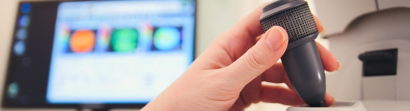
Risposta diretta
A colour Doppler test is an ultrasound technique that maps blood flow inside vessels, the heart or organs using colour to show flow direction and a spectral trace to measure speed. The colour Doppler test helps assess clots, narrowing, valve flow and placental circulation, but it depends heavily on operator skill and does not image vessel walls or tissue as precisely as CT or MR angiography.
Principali takeaways
- Colour Doppler shows blood flow; it does not show all structural disease and is not a substitute for angiography.
- The scan is operator-dependent, and image quality falls with obesity, bowel gas, oedema, casts, wounds and deep vessels.
- A "normal" Doppler report means nothing abnormal was seen in the segments examined, it does not exclude disease elsewhere.
- Different Doppler studies (venous, arterial, carotid, obstetric, renal, scrotal, echo) are distinct examinations with distinct protocols and preparation.
- In India, any Doppler performed in pregnancy is governed by the PC-PNDT Act, 1994; sex determination is a criminal offence.
A colpo d'occhio
| Articolo | Eccezionale |
|---|---|
| Cos'è | Ultrasound-based mapping and measurement of blood flow |
| Radiazione | Nessuno, utilizza onde sonore |
| Durata tipica | Roughly 20-45 minutes for most single-region studies; bilateral limb, whole-abdomen or detailed obstetric studies can take longer |
| Anestesia | Non richiesto |
| Disagio | Mild pressure from the probe; venous studies need firm compression, which can be uncomfortable in a tender limb |
| Preparazione | Varies by body region and by scanning unit |
| Limitazione principale | Operator- and body-habitus dependent; limited in deep, calcified, gas-obscured or heavily oedematous areas |
| Chi lo interpreta | Radiologist, cardiologist or fetal medicine specialist, read with your clinical history |
Conosciuto anche come
- Colour Doppler ultrasound / color Doppler (US spelling)
- Doppler study, Doppler scan, Doppler test
- Duplex scan or triplex scan (greyscale + colour + spectral Doppler)
- Vascular sonography; "USG Doppler"; "Doppler sonography"
- Venous Doppler for clots is often called a DVT scan in Indian practice
- Obstetric Doppler is often called fetal Doppler or "growth scan with Doppler"
- Colour Doppler of the heart is part of the prova di eco (2D echo with colour Doppler)
What a colour Doppler test is
Ordinary greyscale ultrasound shows structure. Doppler ultrasound adds information about movement. When sound waves bounce off moving red blood cells, their frequency shifts. The machine converts that shift into:
- Colour Doppler: a colour overlay showing whether blood is moving towards or away from the probe, and roughly how fast. Red and blue indicate direction relative to the probe, not artery versus vein.
- Spectral (pulsed-wave) Doppler: a waveform and numerical velocities, used to grade narrowing and calculate indices such as the resistive index or umbilical pulsatility index.
- Power Doppler: more sensitive to slow flow but gives no direction information.
Most reports labelled "colour Doppler" are in fact duplex or triplex studies combining all of these with greyscale imaging.
Why a colour Doppler test is done
- Suspected deep vein thrombosis: one-sided leg swelling, calf pain or tenderness.
- Arterial disease of the limbs: claudication, rest pain, non-healing ulcer, cold or discoloured foot.
- Vene varicose: mapping reflux before treatment.
- Carotid and vertebral assessment: after stroke or transient ischaemic attack, or for a neck bruit.
- Gravidanza: umbilical artery, middle cerebral artery and uterine artery Doppler in growth restriction, pre-eclampsia risk or reduced fetal movements.
- Abdominal and renal: portal vein patency in liver disease, transplant kidney perfusion, suspected renal artery stenosis.
- Scrotale: suspected testicular torsion or varicocele.
- Cuore: valve regurgitation and stenosis, shunts (as part of echocardiography).
- Access surveillance: arteriovenous fistula for dialysis.
Chi dovrebbe sottoporsi a questo test?
A colour Doppler test is appropriate when a doctor has a specific question about blood flow. It is not a general health check-up scan. Groups in whom it is commonly indicated include:
- People with sudden painful swelling of one leg or arm, particularly after surgery, immobility, long travel, pregnancy or cancer.
- People with diabetes or long-standing smoking history who have leg pain on walking, a cold foot or a non-healing foot ulcer.
- People who have had a stroke, mini-stroke or transient visual loss, for carotid assessment.
- Pregnant women with suspected fetal growth restriction, hypertension in pregnancy or abnormal fetal movement patterns, when a fetal medicine specialist advises it.
- People with chronic liver disease, portal hypertension, or before and after liver or kidney transplantation.
- Men with acute severe scrotal pain (urgent) or with infertility or scrotal heaviness (routine).
- People on haemodialysis with a poorly functioning fistula.
Routine Doppler screening in people without symptoms or risk factors is generally not recommended, because it produces incidental findings that lead to anxiety and further testing without clear benefit.
What a colour Doppler test cannot detect or exclude
Questa è la parte che la maggior parte dei rapporti non specifica.
- It does not image coronary arteries. Colour Doppler during an echo assesses valves and chamber flow. A normal echo with colour Doppler does not exclude coronary artery disease. Chest pain needs separate evaluation.
- It does not reliably exclude pulmonary embolism. A negative leg venous Doppler does not rule out a clot in the lungs. If breathlessness or chest pain persists, CT pulmonary angiography or another pathway is needed.
- Calf and pelvic veins are limited territory. Isolated calf-vein and iliac-vein thrombi are frequently missed. Many units scan the proximal veins only and repeat the study after several days if suspicion remains.
- Deep abdominal arteries are hard. Renal artery and mesenteric Doppler is often technically incomplete because of bowel gas and body habitus; a normal study does not exclude renal artery stenosis, and CT or MR angiography may be required.
- Heavy calcification blocks sound. In severely calcified arteries, common in diabetes and chronic kidney disease, velocities may be unmeasurable and the degree of narrowing cannot be graded.
- It does not show the vessel wall in the way CT or MR angiography does, and it cannot map a whole arterial tree for surgical planning in one pass.
- It cannot age a clot with certainty or always distinguish acute from chronic thrombus.
- In pregnancy it does not predict outcome with certainty. Normal umbilical artery Doppler reduces but does not remove risk; the test cannot exclude later deterioration.
- It cannot determine fetal sex disclosure in India: and by law this must never be revealed.
- It does not diagnose cancer. Increased vascularity in a lump is suggestive of nothing on its own; tissue diagnosis is separate.
A finding described as "not seen" or "not visualised" means it was not demonstrated in the segments examined under the conditions on the day. That is not the same as excluded. If your symptoms persist or worsen despite a normal Doppler, go back to the doctor who requested it.
Esami correlati ma diversi
| Visita medica | Domanda a cui risponde | Differenza fondamentale |
|---|---|---|
| Greyscale (plain) ultrasound | What does the structure look like? | No flow information |
| Colour Doppler / duplex | Is blood flowing, which way, how fast? | Adds flow; still limited by depth and gas |
| Venous Doppler (compression study) | Is there a clot in the leg or arm veins? | Relies on probe compression, not only colour |
| Arterial Doppler with ABI | Is limb arterial supply reduced? | Pressure index adds objective data; unreliable in calcified vessels |
| Duplex carotideo | Is there narrowing in the neck arteries? | Cannot see intracranial vessels |
| 2D echo with colour Doppler | How are heart valves and chambers working? | Non visualizza le arterie coronarie |
| eco di stress | Does the heart muscle fail under load? | Functional test for ischaemia; resting echo cannot answer this |
| Eco transesofagea | Detailed valve, clot or endocarditis assessment | Semi-invasive, needs sedation and consent |
| Doppler ostetrico | Is placental and fetal circulation adequate? | Separate protocol from routine anomaly or abdominal scan; PC-PNDT regulated |
| CT / MR angiography | Full anatomical map of a vascular territory | Better for planning intervention; CT uses radiation and iodinated contrast |
| Angiografia con catetere | Definitive road map, with option to treat | Invasive, with procedural risk |
| elastografia | Tissue stiffness (e.g. liver fibrosis) | Different physics entirely; not a flow study |
Which test comes first depends on the clinical problem, not on which test is "better". For a swollen leg, venous Doppler is usually the appropriate first test. For claudication, ankle-brachial index and arterial duplex usually come first, with CT or MR angiography if intervention is being planned. For suspected renal artery stenosis in a large adult, cross-sectional angiography is often more productive than Doppler. For chest pain, Doppler-based tests answer only part of the question.
Come preparare
Preparation is an operational matter and differs between scanning units and between machines. The instructions given by the unit performing your scan take precedence over anything written here.
- Limb, carotid and scrotal Doppler: usually no fasting. Wear loose clothes; the limb or neck must be exposed. Avoid heavy moisturiser on the area.
- Abdominal, renal or portal Doppler: many units ask for a period of fasting to reduce bowel gas. The length varies.
- Obstetric Doppler: in later pregnancy a full bladder is usually not required. Early pregnancy scans may ask for one.
- Carry previous scans, angiograms and discharge summaries: comparison changes interpretation more than most patients expect.
- Do not stop anticoagulants, antiplatelets, antihypertensives or any prescribed medicine for a Doppler scan. If someone suggests you should, speak to the prescribing doctor first.
Se hai il diabete e sei in trattamento ipoglicemizzante
Prolonged fasting can cause hypoglycaemia, especially with insulin or sulfonylureas. Tell the unit in advance, ask for an early slot, and ask your treating doctor how to handle that morning's doses. Carry glucose with you.
Se soffri di insufficienza cardiaca, malattia renale cronica in stadio avanzato o restrizione dei liquidi
Do not drink large volumes of water to fill the bladder unless your treating doctor has approved it. Tell the unit about your fluid limit; the scan can usually be adapted.
In gravidanza
Extended fasting is generally avoided. Obstetric Doppler must be performed only on clinical indication, by a registered facility, with the PC-PNDT Form F completed.
Nei bambini
Fasting rules differ substantially for infants and young children and should come from the paediatric team. A parent normally stays in the room.
Anziani e persone con mobilità ridotta
Some studies need standing or prolonged positioning, for example varicose vein mapping. Tell the unit beforehand so support can be arranged.
Cosa succede durante il test?
- You lie on the couch; positioning varies, on the back for abdominal studies, with the leg externally rotated for venous studies, standing or on a tilt table for vein reflux mapping.
- Warm gel is applied. Gel is needed for sound transmission and can feel cool or sticky.
- The sonographer or doctor moves the probe over the region, pressing at intervals. In venous studies the vein is compressed repeatedly to check whether it collapses.
- You may hear a loud "whooshing" or pulsing sound. This is the Doppler signal, and it is expected.
- You may be asked to breathe in, hold your breath, cough, or squeeze the calf, to provoke flow changes.
- The gel is wiped off afterwards. Normal activity can be resumed straight away unless a procedure was performed.
Quanto tempo richiede il processo
- Single-limb venous Doppler: commonly around 20-30 minutes.
- Bilateral limb arterial or venous studies: spesso 40-60 minuti.
- Carotid duplex: usually 20-30 minutes.
- Abdominal or renal Doppler: often 30-45 minutes, longer when bowel gas is troublesome.
- Obstetric Doppler as part of a growth scan: typically 20-40 minutes, longer if the baby is in an awkward position.
Add time for registration, changing and waiting. A difficult study can take considerably longer; this usually reflects technical difficulty, not bad news. Report availability differs between units, and urgent inpatient studies are generally reported faster than routine outpatient ones.
Comprensione del tuo report
A Doppler report usually describes the segments examined, whether flow was present, whether the vein compressed, waveform shape, and velocities. Common terms include:
- Patent / non-compressible: "Patent" means flow was demonstrated. A vein that will not compress under the probe suggests thrombus.
- Echogenic thrombus: visible clot material within the vein.
- Peak systolic velocity (PSV): rises at a narrowing. Ratios between segments matter more than single numbers.
- Monophasic / biphasic / triphasic waveform: describes arterial flow pattern. Loss of the normal multiphasic pattern suggests significant upstream disease, though ageing and stiff vessels also alter it.
- Resistive index (RI): used in renal and transplant work. RI is non-specific and influenced by heart rate, blood pressure and age.
- Riflusso: backward venous flow lasting beyond a threshold, relevant in varicose veins.
- Umbilical artery PI, absent or reversed end-diastolic flow: obstetric findings that are interpreted alongside growth, liquor and cardiotocography, never alone.
Grading terms such as "mild", "moderate" or "significant" narrowing are partly subjective and depend on the operator, the machine settings and the criteria that unit uses. Two units can grade the same vessel differently. Numbers on a report are not a diagnosis. Results vary depending on individual clinical circumstances, and interpretation belongs to the doctor who requested the test.
Quando i risultati possono essere fuorvianti
Findings that can be falsely reassuring (disease under-called)
- Obesity, marked limb oedema, plaster casts, dressings or subcutaneous emphysema reduce signal and can hide thrombus or stenosis.
- Bowel gas can make abdominal arteries unassessable, so disease is simply not seen.
- Dense calcification produces acoustic shadowing; a stenosis behind it can be missed.
- Very slow flow may not be colour-encoded if machine settings are not optimised, and an occluded segment can look patent, or the reverse.
- Isolated distal calf or pelvic-vein clots frequently escape detection.
- Low cardiac output or severe hypotension reduces velocities everywhere and can mask a narrowing.
Findings that can be falsely alarming (disease over-called)
- Aliasing, colour wrapping when the velocity scale is set too low, mimics turbulence and narrowing.
- External compression by a full bladder, a fibroid, a mass or the probe itself can imitate a stenosis.
- Tortuous vessels create high velocities without true narrowing.
- A stenosis in the vessel next door (for example a tight contralateral carotid) raises flow on the other side and can be over-graded.
- Anaemia, fever, pregnancy, hyperthyroidism and arteriovenous fistulas all raise baseline velocities.
- Chronic, old, organised thrombus can be reported as acute DVT if previous scans are not available for comparison.
- Motion, patient talking, or breathing artefacts create spurious colour.
Bring earlier reports. Comparison is often the single most useful thing that prevents both errors.
Rischi e sicurezza
Standard diagnostic colour Doppler
Diagnostic ultrasound uses sound waves, not ionising radiation, and no injection is required for a routine colour Doppler. It is widely considered low-risk when performed for a clinical reason by trained staff. Doppler deposits more acoustic energy than greyscale imaging, so operators use the ALARA principle, as low as reasonably achievable, and keep Doppler exposure time limited, particularly in fetal and neonatal scanning. Minor discomfort from probe pressure is common, and compression of a tender, clotted leg can hurt.
Obstetric and neonatal Doppler
Because of higher acoustic output, pulsed and colour Doppler in pregnancy, especially in the first trimester and over the fetal brain or eye, is used only where clinically indicated, at the lowest settings and shortest duration. Non-medical "keepsake" or entertainment scanning is not appropriate. Neonatal transcranial Doppler follows similar exposure caution.
Ecografia con mezzo di contrasto
This is a different examination. Microbubble contrast agents are injected intravenously and carry a small risk of allergic or hypersensitivity reactions, including rare serious reactions, and require specific consent and resuscitation facilities. Contrast-enhanced ultrasound is not part of a routine colour Doppler.
Ultrasound-guided intervention
Doppler-guided biopsy, aspiration or vascular access carries the risks of the procedure itself, bleeding, infection, injury to nearby structures, and is not comparable in risk to a diagnostic scan.
bandiere rosse
Emergenza immediata: recatevi al pronto soccorso più vicino.
- Sudden breathlessness, chest pain, coughing blood or collapse, especially with a swollen leg, possible pulmonary embolism.
- Sudden face droop, arm weakness or slurred speech, possible stroke.
- A leg or arm that is suddenly cold, pale, numb, painful and pulseless, possible acute arterial occlusion.
- A leg that is grossly swollen, tense and becoming purple or blistered.
- Sudden severe testicular pain, possible torsion; delay risks loss of the testis.
- Severe abdominal or back pain with faintness, possible aneurysm rupture.
- In pregnancy: heavy bleeding, severe headache with visual disturbance, fits, or clearly reduced fetal movements.
In giornata: contatta oggi stesso il tuo medico
- New one-sided calf or thigh swelling, warmth and tenderness.
- A foot ulcer that is spreading, smelly or surrounded by redness.
- Fever with a red, painful limb.
- Rest pain in the foot that wakes you at night.
- A dialysis fistula that has stopped thrilling or buzzing.
- Reduced fetal movements without other emergency features.
Appuntamento di routine
- Aching, heaviness or visible varicose veins that worsen through the day.
- Leg pain on walking that reliably settles with rest.
- Mild long-standing ankle swelling in both legs.
- Scrotal heaviness or infertility assessment.
- Routine follow-up of a previously known and stable finding.
Situazioni speciali
Gravidanza
Obstetric Doppler is performed on clinical indication, commonly for growth restriction, hypertensive disorders or reduced movements. Findings such as raised umbilical artery pulsatility index or absent end-diastolic flow are managed by an obstetrician or fetal medicine specialist together with growth measurements and fetal monitoring. Under the PC-PNDT Act, 1994, the sex of the fetus cannot be disclosed by anyone, under any circumstances.
Bambini
Paediatric Doppler is used for testicular torsion, renal and transplant perfusion, vascular malformations and neonatal brain circulation. Smaller, higher-frequency probes are used and exposure is minimised. Cooperation matters more than preparation; distraction or a feed often helps.
Gli anziani
Stiff, calcified arteries alter waveform shape and can make velocity criteria unreliable. Ankle-brachial index may be falsely high. Toe pressures or cross-sectional angiography are sometimes needed instead.
Diabete
Medial arterial calcification is common and makes both ABI and Doppler grading less dependable. Neuropathy can mask classic claudication, so a painless non-healing ulcer may be the first sign of arterial disease.
Malattia renale cronica e dialisi
Doppler is used for transplant kidney assessment and fistula surveillance. A raised resistive index in a transplant is non-specific and does not by itself indicate rejection. Fluid restriction must be respected when preparation is discussed.
Insufficienza cardiaca
Low cardiac output lowers velocities and can affect interpretation. Lying flat for a prolonged period may be difficult; tell the unit so positioning can be modified.
Obesità
Deeper tissue reduces image quality. A technically limited study should be stated in the report, and an alternative modality may be needed. South Asians can develop metabolic and vascular disease at a lower body mass index than European-origin populations, so a lower BMI does not imply a lower vascular risk.
Colour Doppler in the Indian context
- Legge PC-PNDT del 1994: every ultrasound machine capable of fetal imaging must be registered, Form F must be completed for pregnancy scans, and prenatal sex determination or disclosure is a criminal offence for both the doctor and anyone requesting it. Genetic counselling and record-keeping rules apply to the facility.
- Cardiopatia reumatica remains an important cause of valve disease in India. Colour Doppler during an echo test is central to grading mitral and aortic valve lesions, and often the first objective sign in a young person with breathlessness.
- Diabetes-related peripheral arterial disease and foot ulceration are common; arterial Doppler forms part of limb-salvage assessment, but calcification limits its accuracy in exactly this group.
- Anaemia, including iron deficiency and thalassaemia trait, is widespread and raises baseline blood velocities, which can lead to over-grading of stenosis if the haemoglobin is not taken into account.
- Malattia epatica cronica from viral hepatitis, alcohol and metabolic dysfunction-associated steatotic liver disease (MASLD, formerly NAFLD) is often assessed with portal Doppler for flow direction and collaterals. Note that Doppler assesses flow, not fibrosis; elastography and blood-based scores address fibrosis.
- Terminologia: patients and referring doctors commonly say "USG Doppler", "Doppler sonography" or "DVT scan"; the heart study is usually called the "echo test". These refer to distinct examinations and it is worth confirming which one has been requested on your prescription.
- Data: if you book through an online form or app, the facility must handle your health data under the Digital Personal Data Protection Act, 2023.
Costi e assicurazioni in India
Colour Doppler charges in India vary widely between cities, between government and private facilities, and between single-region and multi-region studies. Rather than quoting a figure, it is more useful to know what drives it.
- Region and number of areas: a single-limb venous study costs less than a bilateral arterial and venous study.
- Tipologia di studio: obstetric Doppler with a growth scan, or an echo with colour Doppler, is priced differently from a limb study.
- Chi lo esegue e lo segnala: consultant radiologist or fetal medicine specialist involvement affects cost.
- Tipo di struttura: standalone diagnostic centre, corporate hospital, teaching hospital or government facility.
- Livello urbano and whether the scan is out-of-hours, bedside or portable.
- Urgency and reporting speed.
- Add-ons: contrast-enhanced ultrasound, additional regions, or a guided procedure.
Assicurazione: outpatient diagnostics are frequently not covered by standard indemnity health policies unless part of an admission or a specific OPD rider. When the Doppler is done during a hospital admission or as part of an approved day-care procedure, it is usually included in the claim. Government schemes such as Ayushman Bharat PM-JAY cover listed packages at empanelled hospitals. Confirm coverage with your insurer or TPA before the scan, and ask the facility for a written estimate. Prices quoted anywhere are indicative only and change; confirm current charges directly with the facility.
Miti e realtà
| Mito | Fatto |
|---|---|
| Colour Doppler uses radiation | It uses sound waves. There is no ionising radiation involved in a diagnostic Doppler scan. |
| Red means artery, blue means vein | Colour indicates direction of flow relative to the probe, and the machine's colour map can be inverted at the press of a button. |
| A normal Doppler means my vessels are fine | It means no abnormality was demonstrated in the segments examined. Symptoms that persist still need review. |
| A normal leg Doppler rules out a clot in the lungs | It does not. Pulmonary embolism requires separate testing. |
| An echo with colour Doppler checks for blockages in the heart | It assesses valves, chambers and pumping. It does not visualise the coronary arteries. |
| Doppler is always better than CT or MRI for blood vessels | They answer different questions. Doppler gives real-time flow; CT and MR angiography give anatomical maps for planning. |
| Doppler in pregnancy can tell the baby's sex | Disclosure of fetal sex is illegal in India under the PC-PNDT Act, 1994, regardless of the scan type. |
| Everyone over a certain age should have a screening Doppler | Routine screening in people without symptoms or risk factors is not generally recommended and can lead to unnecessary follow-up. |
| A "grade" on a Doppler report is an exact measurement | Grading involves operator judgement, machine settings and unit-specific criteria, and can differ between centres. |
Domande frequenti
L'ecografia Doppler a colori è dolorosa?
The scan itself is not painful. You feel gel and probe pressure. Venous studies require firm, repeated compression of the limb, which can be genuinely uncomfortable if the leg is swollen or tender. Tell the operator if it hurts; pressure can usually be adjusted without compromising the study.
Do I need to fast before a colour Doppler?
Fasting is usually asked for abdominal, renal or portal Doppler to reduce bowel gas, and is generally not needed for limb, carotid or scrotal studies. Fasting duration differs between units. If you take insulin or other glucose-lowering medicines, ask your doctor before fasting and request an early appointment.
How is a colour Doppler different from a normal ultrasound?
A plain ultrasound shows structure in greyscale. Colour Doppler adds information about blood movement, direction, speed and pattern, overlaid in colour, plus waveform measurements. Most Doppler studies combine both. The extra information helps with clots, narrowing and organ perfusion, but does not improve structural detail.
Can a colour Doppler detect a blocked heart artery?
No. Colour Doppler during echocardiography assesses valves, shunts and chamber flow. It does not visualise the coronary arteries. Suspected coronary disease needs a different pathway, such as an ECG, treadmill test, stress imaging, CT coronary angiography or invasive angiography, as your cardiologist advises.
Is colour Doppler safe in pregnancy?
Diagnostic ultrasound in pregnancy is widely used and considered low-risk when clinically indicated. Doppler delivers more acoustic energy than plain imaging, so it is used selectively, at minimum settings and for the shortest time, particularly in early pregnancy. Non-medical keepsake scanning is not appropriate.
Why did my report say the study was "technically limited"?
This means image quality was reduced, commonly by bowel gas, body habitus, oedema, dressings, calcification or patient movement. It is a statement of honesty, not an error. It means findings are less certain, and your doctor may repeat the scan later or choose a different test.
Can I drive and work after the scan?
Yes, after a routine diagnostic colour Doppler you can drive and return to normal activities immediately. The situation differs if you also had contrast-enhanced ultrasound, sedation, a transoesophageal echo or a guided procedure, in those cases follow the specific discharge instructions given.
How often should a colour Doppler be repeated?
There is no fixed interval. Repeat frequency depends on the condition, for example, surveillance after DVT, monitoring a dialysis fistula, or serial obstetric Doppler in growth restriction, which may be every few days. Your treating doctor sets the interval based on your clinical situation.
My Doppler is normal but the symptoms continue. What now?
Return to the doctor who requested the test. A normal Doppler reports what was seen in specific segments on that day; it does not exclude disease elsewhere or disease that develops later. Your doctor may repeat the scan, examine a different territory, or move to CT or MR angiography.
fonti
- Pre-Conception and Pre-Natal Diagnostic Techniques (Prohibition of Sex Selection) Act, 1994, Government of India, with subsequent rules and amendments.
- British Medical Ultrasound Society, safety guidelines for the safe use of diagnostic ultrasound equipment.
- World Federation for Ultrasound in Medicine and Biology, statements on the safe use of Doppler ultrasound, including fetal applications.
- International Society of Ul











 Il miglior ospedale vicino a me Chennai


