


Izravan odgovor
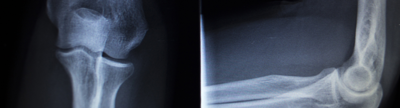
An elbow X-ray is a quick imaging test that uses a small dose of ionising radiation to show the bones and joint spaces of the elbow. The elbow X-ray is mainly used to look for fractures, dislocations and arthritic change. An elbow X-ray does not show ligaments, tendons, nerves or cartilage directly.
Ključni postupci
- Bones, not soft tissue: The elbow X-ray images bone and joint alignment, not ligaments, tendons or nerves.
- Normal does not mean nothing wrong: Some fractures, especially in children and older adults, are not visible on the first X-ray.
- Brzi test: Image acquisition usually takes a few minutes; the whole visit is commonly 10-20 minutes depending on how busy the unit is.
- Other tests answer other questions: MRI, CT and ultrasound each show things the elbow X-ray cannot.
- Interpretacija je klinička: The doctor who requested the elbow X-ray reads the report alongside your symptoms and examination. Results vary depending on individual clinical circumstances.
Na prvi pogled
| svojstvo | Detalji |
|---|---|
| Što je to slika | Bones of the elbow: distal humerus, proximal radius and ulna, joint spaces and alignment |
| Standardni prikazi | Anteroposterior (AP) and lateral; oblique or axial views added when needed |
| Radijacija | Low dose ionising radiation; limb X-rays are among the lower-dose radiographic studies |
| Priprema | Usually none beyond removing metal from the arm; follow your unit's instructions |
| Vrijeme provedeno u rendgenskoj sobi | Typically a few minutes of positioning and exposure |
| nelagodnost | Positioning an injured elbow can hurt; the exposure itself is not felt |
| Ne može se prikazati | Ligament tears, tendon disease, nerve compression, cartilage detail, most occult fractures |
Također poznat kao
- Elbow radiograph
- X-ray elbow AP and lateral
- Elbow film
- Kohni ka X-ray (common Hindi phrasing used by patients)
- Right elbow / left elbow X-ray (side is always specified on the request)
What an elbow X-ray is
An elbow X-ray passes a controlled beam of X-rays through the joint onto a digital detector. Dense structures such as bone absorb more of the beam and appear white; muscle, fat and fluid absorb less and appear in shades of grey and black. Ligaments and tendons are not distinguished from surrounding soft tissue on a plain radiograph.
A radiographer positions the arm for each view. The AP view is taken with the elbow straightened and the palm facing up where the injury allows; the lateral view is taken with the elbow bent, usually to about 90 degrees. If the elbow cannot be straightened because of pain or a plaster, modified views are used and the report will usually say the views were limited.
Why an elbow X-ray is done
- Suspected fracture after a fall on an outstretched hand, direct blow or road traffic injury.
- Suspected dislocation or to confirm position after a joint has been reduced.
- Arthritic change: joint space narrowing, osteophytes (bone spurs), subchondral change.
- Opuštena tijela or calcified fragments within the joint.
- Bone lesions: to look for a destructive or expansile bone abnormality when symptoms or examination raise that concern.
- Strano tijelo that is radio-opaque, such as metal or glass, after a penetrating injury.
- Checking healing or hardware after fracture treatment, plating or joint replacement.
- Procjena deformiteta in malunion or after childhood injury.
Who should get an elbow X-ray
An elbow X-ray is usually appropriate for:
- People with elbow pain and swelling after a definite injury, particularly if they cannot fully straighten the elbow, cannot bear weight through the arm, or have visible deformity.
- Children with elbow injury and localised bony tenderness, paediatric elbow fractures are common and easy to miss clinically.
- Older adults after a fall, where bone may be fragile.
- People with longstanding elbow stiffness, locking or grating suggesting arthritis or loose bodies.
- People with suspected infection in or around the joint, as part of a wider assessment.
- Follow-up of a known fracture, surgical fixation or implant.
An elbow X-ray is often ne the test that answers the question in tennis elbow (lateral epicondylitis), golfer's elbow, cubital tunnel syndrome or simple soft-tissue bruising. In those situations the diagnosis is usually clinical, and imaging is chosen based on what needs to be clarified.
What an elbow X-ray cannot detect or exclude
This is the most important limitation to understand before reading a report.
- Ozljede ligamenata: A tear of the ulnar (medial) collateral ligament or lateral ligament complex is not visible. A normal elbow X-ray does not exclude ligament rupture or instability.
- Tendon problems: Tennis elbow, golfer's elbow and distal biceps or triceps tendon tears are not diagnosed on a plain film. Sometimes calcification near the epicondyle is seen, but its presence or absence does not confirm or rule out tendinopathy.
- Nerve problems: Ulnar nerve compression at the cubital tunnel and radial nerve entrapment are not visible.
- Hrskavica: Early cartilage damage and small osteochondral lesions are frequently not apparent. Joint space on X-ray is an indirect surrogate for cartilage thickness.
- Occult and undisplaced fractures: Hairline radial head fractures, some supracondylar and condylar fractures in children, and stress fractures may not show on the initial films. A negative elbow X-ray does not exclude a fracture.
- Bone marrow oedema and early infection: Osteomyelitis often shows no radiographic change in the first one to two weeks. Early septic arthritis may look normal.
- Muscle, bursa and fluid detail: Olecranon bursitis, haematoma and abscess are soft-tissue diagnoses; X-ray may show only indirect displacement of fat planes.
- Strana tijela neprozirna za radiografiju: Wood, most plastics and many thorns do not show.
A finding described as "not identified" or "not seen" means it was not visible on those images. It does not mean the problem has been excluded. If your elbow symptoms persist, worsen or change despite a normal X-ray, go back to your doctor, repeat films after 7-14 days, or a different test altogether, may be needed.
Povezani, ali različiti pregledi
These tests are not ranked from worst to best. Each answers a different question, and the right first test depends on the clinical problem.
| test | Pitanje na koje odgovara | Glavno ograničenje |
|---|---|---|
| Elbow X-ray (radiograph) | Is there a fracture, dislocation, arthritic change, loose calcified body or bone lesion? | Blind to ligaments, tendons, nerves, cartilage detail; misses occult fractures |
| CT elbow | Exact bone anatomy in complex or comminuted fractures; surgical planning; fracture suspected despite normal X-ray | Higher radiation dose than a limb radiograph; limited soft-tissue and ligament detail |
| MRI elbow | Ligament tears, tendon disease, cartilage and osteochondral lesions, bone marrow oedema, occult fracture, early infection, soft-tissue masses | Costlier, longer, less available; metal implants require MRI safety assessment; motion degrades images |
| Ultrasound elbow | Tendon and bursal problems, joint effusion, guided injection or aspiration, ulnar nerve at the cubital tunnel | Cannot see inside bone; operator-dependent; deep structures less well seen |
| Elbow arthrogram or MR arthrogram | Intra-articular loose bodies and cartilage surfaces when plain MRI is inconclusive | Invasive contrast injection; specialised availability |
| Nerve conduction study / EMG | Whether and where a nerve is being compressed or damaged | Not an imaging test; does not show structure |
A plain elbow X-ray and an ultrasound-guided elbow injection are different procedures even though both involve imaging. Similarly, a diagnostic elbow radiograph is not the same as a post-reduction check film or a series taken under image intensifier in theatre.
How to prepare for an elbow X-ray
Preparation is simple, but the exact instructions are operational and set by the unit performing the study. Your imaging department's instructions take precedence over anything you read here.
- Odjeća: Wear a short-sleeved or loose-sleeved top so the arm can be exposed. A gown may be offered.
- Metal: Remove watches, bangles, bracelets, rings and any metal on the arm. Metal creates dense shadows that can obscure bone.
- Bez posta: A plain elbow X-ray needs no fasting, no injection and no contrast. You can eat, drink and take your usual medicines as prescribed.
- Bring the request and prior films: Previous elbow X-rays help the radiologist judge whether a change is new.
- Tell the radiographer: If you are or might be pregnant, if the arm is in a plaster or splint, if you have implants or previous surgery in the arm, or if pain prevents certain positions.
Ako ste trudni ili biste mogli biti trudni
Tell the radiographer before the exposure. An elbow is far from the uterus and is shielded and collimated, so foetal dose from a limb radiograph is extremely low. Even so, the department will confirm the study is needed and may offer abdominal shielding. Pregnancy is not an automatic reason to avoid a necessary limb X-ray.
Djeca
A parent or carer is usually allowed to stay, wearing a lead apron, to help the child keep still. Comparison views of the uninjured elbow are sometimes requested because growth plates can be mistaken for fractures, that decision rests with the radiologist or treating doctor. Paediatric exposure settings are lower than for adults.
If your arm is in a plaster or splint
Films can be taken through plaster. The images will be less clear, and the report may note this. Do not remove a splint yourself; ask the treating team.
Limited mobility or severe pain
Tell the staff if you cannot straighten or bend the elbow. Modified or seated positions can be used. Pain relief timing should be discussed with the treating doctor.
What happens during an elbow X-ray
The sequence is short and predictable:
- Identification and checks: Your details, the side to be imaged and pregnancy status where relevant are confirmed.
- pozicioniranje: The arm is rested on the detector or table. For the AP view the elbow is as straight as possible; for the lateral view it is bent with the thumb pointing up.
- Izlaganje: You hold still and breathe normally for a fraction of a second per image. The radiographer steps behind a screen. You will not feel the X-ray.
- Dodatni prikazi: Oblique or specialised views may be added if the standard two views do not answer the question.
- Provjera slike: The radiographer reviews the images for quality and may repeat a view.
- Nakon: Nothing is injected and there is no recovery period. You may leave and return to normal activity, subject to any restriction advised for the injury itself.
Koliko dugo traje proces
- Actual exposures: milliseconds each.
- Time in the X-ray room: usually around 5 minutes, longer if the elbow is painful, plastered or needs extra views.
- Ukupan posjet: commonly 10-20 minutes including registration, positioning and image checks; longer at busy times.
- Izvješće: in an emergency the treating doctor often looks at the images immediately. A formal radiologist report follows separately, and reporting times differ between units, ask the department when and how you will receive it.
Understanding your elbow X-ray report
Reports describe what was seen on the images, in radiological language. Common terms include:
- No acute bony injury / no fracture identified: No break was visible on these views. This is a description, not a guarantee that no fracture exists.
- Joint spaces preserved: The gaps between bones look normal in width, which indirectly suggests reasonable cartilage thickness.
- Joint space narrowing, osteophytes, subchondral sclerosis: Features often seen in osteoarthritis. Radiographic changes correlate imperfectly with pain and function, some people with marked changes have few symptoms, and vice versa.
- Elevated fat pads / positive posterior fat pad sign: Suggests fluid or blood in the joint, which raises the possibility of a fracture that is not directly visible and often prompts further assessment.
- Izljev: Fluid in the joint. X-ray cannot tell whether that fluid is blood, inflammatory fluid or pus.
- Loose body / intra-articular calcification: A calcified fragment within the joint. Cartilage-only fragments will not show.
- Displacement, angulation, comminution: Describes how fracture fragments sit; these terms guide treatment.
- Periarticular calcification: Calcium near the joint, which may be old injury, tendon calcification or other causes.
- Limited views / suboptimal positioning: The study could not be performed ideally, so sensitivity is reduced.
No radiographic term is a diagnosis on its own. The doctor who requested the elbow X-ray combines the report with your history, examination and, where needed, further tests.
When elbow X-ray results can be misleading
Falsely reassuring: appears normal when a problem exists
- Undisplaced or hairline fractures: Radial head and neck fractures can be invisible initially and only become apparent on repeat films after 10-14 days.
- Paediatric fractures at growth plates: Cartilage is radiolucent, so Salter-Harris type I injuries and some condylar fractures can look normal.
- Early osteomyelitis or septic arthritis: Bone changes lag behind infection by one to two weeks.
- Soft-tissue and ligament injury: Complete ligament rupture with instability may show a perfectly normal-looking bone film.
- Poor positioning or plaster overlay: A true lateral view is needed to judge subtle fracture lines and the fat pad signs; a rotated film can hide them.
- Osteopenija: In fragile bone, fracture lines can be indistinct.
Falsely alarming: appears abnormal when it is not clinically significant
- Normal growth plates and ossification centres in children can be mistaken for fracture fragments; the elbow has six centres that appear and fuse at different ages.
- Accessory ossicles and normal variants may resemble loose bodies.
- Old, healed injury: Deformity or calcification from a childhood injury can be reported as an abnormality although it is not the cause of current pain.
- Age-related degenerative change is common and may be incidental rather than the source of symptoms.
- Overlapping metal, plaster or clothing artefact can mimic a lesion.
Rizici i sigurnost
Plain elbow radiograph
A limb X-ray uses a low dose of ionising radiation, among the lower doses in diagnostic radiography, and the elbow is distant from radiosensitive organs. There is no injection and no recovery period. Radiation risk is not zero, which is why X-rays are requested only when the answer will change management, and why departments use collimation, shielding and the lowest practical exposure. Repeated films over time add up, so tell your doctor about recent imaging of the same area.
Repeat and follow-up films
Follow-up radiographs during fracture healing mean more than one exposure. This is usually justified by the need to check alignment and union, but each film should have a clear purpose.
CT and MRI of the elbow: different risk profiles
CT uses a higher radiation dose than a plain elbow film. MRI uses no ionising radiation, but pacemakers, neurostimulators, cochlear implants, metallic fragments and some orthopaedic hardware require MRI safety assessment before scanning. Contrast-enhanced studies carry additional considerations, including allergy and kidney function, which the requesting team will assess.
nelagodnost
The main discomfort is moving an injured elbow into position. Tell the radiographer if a position is unbearable, an alternative view can usually be used.
Red flags: when to seek help rather than wait for a scan or report
Hitna pomoć: odmah idite na hitnu pomoć
- Obvious deformity of the elbow, or the joint looks dislocated.
- Bone visible through a wound, or a deep wound over the joint.
- Hand or forearm that is cold, pale, blue or pulseless, or severe pain out of proportion to the injury with a tight, tense forearm.
- Numbness, tingling or weakness of the hand that started after the injury, or inability to move the fingers or wrist.
- Rapidly increasing swelling after injury, especially if you take blood thinners.
- Fever with a hot, red, extremely painful elbow that you cannot move.
Same day: contact your doctor or an urgent care service today
- Inability to straighten or fully bend the elbow after an injury.
- Significant swelling and pain that prevent using the arm.
- A child refusing to use the arm after a fall.
- Spreading redness, discharge from a wound, or fever after elbow surgery or injection.
- Pain that worsens rather than settles over the first 24-48 hours after injury.
Rutinski pregled
- Elbow pain that persists for weeks despite rest, with or without a normal X-ray.
- Gradual stiffness, locking, clicking or catching.
- Mild aching with activity, or suspected tennis or golfer's elbow.
- Follow-up review of a healing fracture as scheduled by your treating team.
Posebne situacije
Djeca i adolescenti
The growing elbow is one of the trickiest areas in paediatric radiology. Ossification centres appear in a predictable sequence and can mimic or mask injury. Supracondylar fractures are the commonest elbow fracture in children and may be subtle. If a child remains tender and will not use the arm despite a normal X-ray, ask for review, clinical assessment outweighs a normal film.
Trudnoća
A clinically necessary elbow X-ray is generally performed during pregnancy with shielding and tight collimation, as foetal dose from a limb study is very low. Always declare pregnancy so the team can document and optimise the study.
Starije odrasle osobe
Bone may be fragile, so relatively minor falls can cause fractures. Osteopenia makes fracture lines harder to see. Distal humerus and radial head fractures in this group sometimes need CT for surgical planning. A fall itself also deserves assessment for its cause.
People with metal implants or previous elbow surgery
Plates, screws and joint replacements are clearly visible and X-ray remains the main tool for checking hardware position, loosening and union. Tell the radiographer about implants, and mention them again if MRI is later suggested, as MRI safety assessment will be required.
People with diabetes, chronic kidney disease or immunosuppression
These groups are at higher risk of joint and bone infection, which plain X-rays may not show early. If there is fever, spreading redness or a non-healing wound, further tests and blood investigations are usually needed regardless of a normal elbow X-ray. A plain X-ray itself requires no change to any medicine.
People with reduced ability to hold still
Tremor, dementia, spasticity or severe pain can blur images. Support pads, shorter exposure settings and an accompanying carer help. Repeat exposures are sometimes unavoidable.
Elbow X-ray in the Indian context
- Access and terminology: Plain radiography is the most widely available imaging service across India, from district hospitals to small diagnostic centres. Patients commonly ask for an "X-ray of kohni" or "elbow film", and requests are usually written as "X-ray elbow AP and lateral".
- Uobičajene prezentacije: Road traffic injuries, falls from height, sports and playground injuries, and agricultural or industrial machinery injuries are frequent reasons for elbow radiographs. Cricket and other throwing sports contribute to medial elbow problems, where X-ray is often normal and clinical assessment or MRI is what clarifies matters.
- Tuberkuloza: Osteoarticular tuberculosis remains a relevant differential in India for a chronically swollen, stiff, painful joint with bone destruction on X-ray. Radiographic features can overlap with other infections and with inflammatory arthritis, so diagnosis usually needs further imaging, aspiration or biopsy and microbiological testing rather than the X-ray alone.
- Traditional bone-setting: Manipulation by untrained bone-setters before medical review is still seen in parts of the country and can be associated with malunion, stiffness or missed injury. If an elbow injury has already been manipulated, tell the doctor, as X-ray findings must be interpreted in that light.
- Digital reports: Many centres now provide digital images and reports by link or application. Personal health data shared through such portals and forms is covered by the Digital Personal Data Protection Act 2023; ask how your images and reports are stored and shared.
- Radiation safety oversight: X-ray installations in India operate under Atomic Energy Regulatory Board requirements for equipment registration, room shielding and staff monitoring. It is reasonable to ask a centre about lead shielding for accompanying relatives.
Troškovi i osiguranje u Indiji
An elbow X-ray is one of the lower-cost imaging tests in India. Prices vary widely between government facilities, standalone diagnostic centres and corporate hospitals, and between cities and smaller towns, so ask the specific centre for their current rate rather than relying on a published figure.
Čimbenici koji utječu na to koliko plaćate:
- Number of views: Two views cost more than one; oblique or comparison views of the other elbow add to the total.
- Postavljanje: Emergency department imaging, bedside portable films and after-hours studies usually cost more than a scheduled outpatient X-ray.
- Digital versus film: Hard-copy film, CD or printed reports may be charged separately.
- Izvješćivanje: Some centres bundle the radiologist report; others charge for it, or for an urgent report.
- Follow-up films: Fracture care typically involves several radiographs over weeks.
- eskalacija: If CT or MRI is subsequently needed, costs rise substantially.
Osiguranje: Outpatient diagnostic tests are often not covered by standard indemnity health insurance unless the policy includes an OPD or diagnostic benefit. Imaging done as part of an admission, or in the pre- and post-hospitalisation window for a covered claim, is more commonly reimbursed. Accident and personal injury policies, employer schemes, government schemes such as Ayushman Bharat PM-JAY at empanelled facilities, CGHS and ESIC each have their own rules. Confirm coverage, cashless eligibility and documentation with your insurer or the hospital insurance desk before the test where possible.
Mitovi i činjenice
| Mit | Činjenica |
|---|---|
| "A normal X-ray means my elbow is fine." | A normal radiograph means no abnormality was visible on those views. Ligament, tendon, nerve and cartilage injuries, and some fractures, can be present with a normal film. |
| "X-rays show tennis elbow." | Tennis elbow is a tendon problem diagnosed clinically. X-ray is used mainly to look for other causes of pain, not to confirm it. |
| "An X-ray gives a dangerous dose of radiation." | A limb X-ray uses a low dose and the elbow is away from radiosensitive organs. Risk is small but not zero, so exposures are kept to what is clinically needed. |
| "MRI je uvijek bolja od rendgena." | MRI and X-ray answer different questions. For fracture, alignment and implant assessment, radiographs are often more practical and sufficient. |
| "X-rays cannot be done in pregnancy." | A necessary limb X-ray is usually performed with shielding, as foetal dose is very low. Declare pregnancy so the team can optimise the study. |
| "The bone-setter fixed it, so no X-ray is needed." | Manipulation without imaging can leave fractures malaligned or unrecognised. Medical assessment with appropriate imaging is advisable after any significant elbow injury. |
| "I can read my own X-ray online." | Interpreting elbow films, especially in children, requires training. Findings must be matched to your symptoms and examination by your doctor. |
Često postavljana pitanja
Does an elbow X-ray hurt?
The X-ray exposure itself is not felt. What can hurt is straightening or bending an injured elbow for the standard views. Tell the radiographer if a position is too painful; modified views are available. Nothing is injected, and there is no recovery time afterwards.
Can an elbow X-ray miss a fracture?
Yes. Undisplaced radial head fractures, some paediatric growth-plate injuries and stress fractures can be invisible on the first films. If tenderness and loss of movement persist, doctors may repeat the X-ray after 10-14 days or arrange CT or MRI. Persisting symptoms always warrant review.
How many X-ray views of the elbow are usually taken?
Two views, anteroposterior and lateral, are standard. Oblique, axial or radial-head-specific views may be added when the standard views do not answer the clinical question. Comparison views of the uninjured elbow are occasionally requested in children, at the radiologist's or treating doctor's discretion.
Will an elbow X-ray show a ligament tear or nerve problem?
No. Ligaments, tendons and nerves are not visualised on a plain radiograph. Suspected collateral ligament tears or distal biceps rupture are typically assessed with MRI or ultrasound, and nerve compression with clinical examination plus nerve conduction studies where needed.
Do I need to fast or stop my medicines before an elbow X-ray?
No fasting is required and a plain elbow X-ray needs no contrast or injection. Continue prescribed medicines as advised by your prescriber; do not stop or change anything for this test. Follow any specific instructions your imaging unit gives you.
How soon will I get the elbow X-ray report?
In emergency and orthopaedic settings, the treating doctor often reviews the images straight away. A formal radiologist report is issued separately, and timing differs between facilities. Ask the imaging department when the report will be ready and how you will receive it.
Is an elbow X-ray safe for children?
Paediatric elbow X-rays are performed frequently and use reduced exposure settings with collimation and shielding. A carer can usually stay, wearing a lead apron. The benefit of identifying a fracture in a child who will not use the arm generally outweighs the small radiation risk.
What is a positive fat pad sign on my report?
An elevated or visible posterior fat pad suggests fluid or blood within the elbow joint. It does not identify a fracture directly, but it raises the possibility of one that is not visible, so your doctor may treat you as if a fracture is present and repeat imaging.
Can an elbow X-ray diagnose arthritis?
An elbow X-ray can show features associated with osteoarthritis, such as joint space narrowing and osteophytes, and some changes seen in inflammatory arthritis. It cannot tell which type of arthritis you have on its own; blood tests and clinical assessment are needed for that.
Izvori
- Royal College of Radiologists, iRefer: Making the Best Use of Clinical Radiology, referral guidance on musculoskeletal radiography.
- American College of Radiology Appropriateness Criteria, Acute Elbow and Forearm Pain; Chronic Elbow Pain.
- International Commission on Radiological Protection guidance on justification and optimisation in diagnostic radiology.
- Atomic Energy Regulatory Board (India), safety requirements for medical diagnostic X-ray installations.
- Zakon o zaštiti digitalnih osobnih podataka iz 2023. (Indija), rukovanje osobnim zdravstvenim podacima.
- Standard orthopaedic and radiology texts on elbow trauma, paediatric elbow ossification and osteoarticular tuberculosis.









 Najbolja bolnica u mojoj blizini Chennai


