


સીધો જવાબ
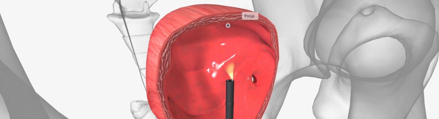
Cystoscopy is a procedure in which a urologist passes a thin telescope (cystoscope) through the urethra to view the bladder lining and urethra directly. Cystoscopy is used mainly to investigate blood in urine, recurrent infection, bladder tumours and voiding problems. Cystoscopy examines the lower urinary tract only, it does not image the kidneys.
કી ટેકવેઝ
- What cystoscopy sees: the inside surface of the urethra, prostatic urethra in men, and bladder lining.
- What cystoscopy misses: kidney and ureteric disease, tumours within the bladder wall or outside it, and flat lesions that can be subtle on white-light viewing.
- Two main forms: flexible cystoscopy (awake, local anaesthetic gel) and rigid cystoscopy (usually under spinal or general anaesthesia, allows fuller therapeutic work).
- Time involved: a straightforward diagnostic flexible cystoscopy commonly takes about 5-15 minutes of scope time; rigid or therapeutic procedures take longer.
- A normal cystoscopy is reassuring but not absolute. If symptoms such as visible blood in urine continue, go back to your doctor, further imaging or repeat evaluation may be needed.
એક નજરમાં
| વસ્તુ | વિગતવાર |
|---|---|
| તપાસાયેલ વિસ્તાર | Urethra and bladder (lower urinary tract) |
| તપાસ કરી નથી | Kidneys, ureters (unless a separate ureteroscopy or retrograde study is added) |
| પ્રકાર | Flexible diagnostic; rigid diagnostic/therapeutic; with biopsy or resection; blue-light or narrow-band imaging where available |
| એનેસ્થેસીયા | Local lignocaine gel for flexible; spinal or general for most rigid procedures |
| Typical scope time | About 5-15 minutes diagnostic; longer if biopsy, stone removal or resection is done |
| પુનઃપ્રાપ્તિ | Most people go home the same day after flexible cystoscopy; burning and pink urine for 1-2 days is common |
| કોણ કરે છે | યુરોલોજિસ્ટ |
તરીકે પણ જાણીતી
- સિસ્ટોરેથ્રોસ્કોપી
- Bladder endoscopy / bladder telescopy
- "Bladder camera test" or "bladder scopy" in common Indian usage
- Check cystoscopy, the surveillance examination done after bladder tumour treatment
- TURBT, transurethral resection of bladder tumour, a therapeutic operation performed through a rigid resectoscope, not the same as diagnostic cystoscopy
What cystoscopy is
Cystoscopy uses a lighted telescope with a camera, passed along the urethra into the bladder. Sterile fluid (usually saline) distends the bladder so the lining can be seen clearly. Images appear on a monitor in real time.
A flexible cystoscope bends, is narrow, and is normally used awake in a procedure room with anaesthetic gel. A rigid cystoscope is straight, has wider working channels, and allows instruments for biopsy, stone fragmentation, resection of tumours, stent placement or removal; rigid work is usually done in an operating theatre under spinal or general anaesthesia.
Cystoscopy may be purely diagnostic (look only) or therapeutic (look and treat). The plan is decided by the urologist based on the reason for the examination.
Why cystoscopy is done
- પેશાબમાં લોહી: visible (gross) haematuria, and in selected people microscopic haematuria, especially with risk factors such as smoking or age above 40-45.
- Suspected or known bladder cancer: for initial assessment and for surveillance after treatment of non-muscle-invasive bladder cancer.
- વારંવાર પેશાબની નળીઓનો વિસ્તાર ચેપ that does not fit the usual pattern, or infection with unusual organisms.
- Bladder stones or foreign bodies, including retained stents or migrated devices.
- Obstructive voiding symptoms: assessment of urethral stricture or prostatic obstruction before surgery.
- Bladder pain syndrome / interstitial cystitis: often as cystoscopy with hydrodistension under anaesthesia.
- Persistent irritative symptoms (urgency, frequency, pain) with no explanation on urine tests and imaging.
- Procedural access: ureteric stent insertion or removal, botulinum toxin injection into the bladder, urethral dilatation.
આ ટેસ્ટ કોણે કરાવવો જોઈએ?
Cystoscopy is not a screening test for the general population. It is generally considered for:
- Adults with even one episode of painless visible blood in urine, once other causes have been considered.
- Adults with microscopic haematuria that persists after treatment of an infection, particularly with smoking history, occupational exposure to aromatic amines (dye, rubber, leather, paint industries) or age over 40.
- People with a previous bladder tumour, on a surveillance schedule set by their urologist.
- Men with poor urinary flow when a stricture is suspected, or before transurethral prostate surgery in selected cases.
- Women with recurrent UTI where an anatomical cause, stone or fistula is suspected.
- People with suspected bladder stone, retained stent or bladder foreign body.
Cystoscopy is usually deferred during an active, untreated urinary infection, except when it is being done as an emergency. Whether cystoscopy is appropriate for you is a clinical decision made by the treating urologist.
What cystoscopy cannot detect or exclude
- Kidney and ureteric disease. Cystoscopy does not visualise the kidneys or most of the ureters. Upper tract tumours, kidney stones and hydronephrosis need CT urography, ultrasound or retrograde/ureteroscopic study. In haematuria work-up, cystoscopy and upper tract imaging are complementary, neither replaces the other.
- Disease within or beyond the bladder wall. Cystoscopy shows the inner lining. Depth of tumour invasion, lymph nodes and spread require biopsy, resection specimens and cross-sectional imaging (CT/MRI).
- Flat, subtle lesions. Carcinoma in situ can appear as mild redness or be missed on standard white-light viewing. Enhanced techniques (blue-light or narrow-band imaging) and urine cytology are used in selected cases; availability varies.
- Bladder function. Cystoscopy shows structure, not how the bladder stores and empties. Urodynamic studies and uroflowmetry answer functional questions.
- પ્રોસ્ટેટ કેન્સર. Cystoscopy is not a prostate cancer test. It shows the shape of the prostatic urethra, not cancer within the gland.
- Infection organism. Cystoscopy cannot identify which bacterium is present, urine culture does that.
A cystoscopy reported as normal means no abnormality was seen on that examination. That is not the same as disease being excluded. If blood in urine, pain or urinary symptoms continue after a normal cystoscopy, return to your doctor rather than assuming the matter is closed.
સંબંધિત પરંતુ અલગ પરીક્ષાઓ
| પરીક્ષા | પ્રશ્ન એ જવાબ આપે છે | Key difference from cystoscopy |
|---|---|---|
| લવચીક સિસ્ટોસ્કોપી | Is the bladder lining and urethra normal? | Awake, diagnostic, limited therapeutic capability |
| Rigid cystoscopy / TURBT | Can the lesion be biopsied or resected now? | Anaesthesia needed; wider instruments; operative |
| Ureteroscopy (URS / RIRS) | What is inside the ureter or kidney? | Scope passed beyond the bladder into the upper tract |
| Ultrasound KUB (USG/sonography) | Hydronephrosis, kidney stones, post-void residue, larger bladder masses | Non-invasive; misses small and flat bladder lesions; cannot biopsy |
| સીટી યુરોગ્રાફી | Upper tract tumours and stones, ureteric anatomy | Radiation and usually iodinated contrast; poorer for flat bladder lesions |
| પેશાબની સાયટોલોજી | Are malignant cells being shed? | Laboratory test; good for high-grade disease, weak for low-grade tumours |
| Urodynamics / uroflowmetry | How does the bladder store and empty? | Functional, not visual |
| Cystogram / MCU | Reflux, leaks, fistula, bladder outline | X-ray contrast study; no direct view of the lining |
These tests answer different questions. Which comes first depends on the presentation: for painless visible haematuria in an adult, cystoscopy plus upper tract imaging are typically both required; for suspected acute ureteric colic, imaging comes first and cystoscopy has no primary role; for suspected recurrent UTI, urine culture and ultrasound usually precede cystoscopy.
કેવી રીતે તૈયાર કરવું
Preparation instructions are set by the unit performing the procedure and vary with the type of cystoscopy and anaesthesia planned. Your unit's written instructions take precedence over anything here.
- પેશાબ પરીક્ષણ: many units check a urine sample beforehand; an active untreated infection often means postponing.
- ઉપવાસ: generally not required for awake flexible cystoscopy. Fasting is required when spinal or general anaesthesia is planned; the anaesthesia team will specify the duration.
- દવાઓ: tell the team about blood thinners (aspirin, clopidogrel, warfarin, newer oral anticoagulants), diabetes medicines, and any allergy to lignocaine, latex or antiseptics. Do not stop or change any prescribed medicine yourself, only the prescribing doctor or the urology team should advise this.
- Bladder fullness: instructions differ. Some units ask you to arrive with a comfortably full bladder; many ask you to empty the bladder immediately before a flexible cystoscopy. Follow your unit.
- એન્ટીબાયોટિક્સ: a single preventive dose is given in some situations, not routinely in all. This is decided case by case.
- પરિવહન: arrange someone to accompany you home if sedation or anaesthesia is used.
જો તમને ગ્લુકોઝ ઘટાડતી સારવાર પર ડાયાબિટીસ હોય તો
Fasting for anaesthesia can cause low blood sugar. Ask the team in advance how to handle insulin or tablets on the day, and carry a glucose source. Certain diabetes medicines are stopped before surgery under medical instruction only.
જો તમને ક્રોનિક કિડની રોગ, હૃદયની નિષ્ફળતા અથવા પ્રવાહી પ્રતિબંધ હોય
General advice to "drink plenty of fluids" is not safe for everyone. If you are on a fluid restriction or diuretics, ask the treating team for a specific fluid plan before and after the procedure.
ગર્ભાવસ્થામાં
Cystoscopy can be performed in pregnancy when clearly indicated, for example for stent placement in obstruction. Timing, positioning and anaesthesia are modified. Tell the team if you are or may be pregnant.
બાળકો
Paediatric cystoscopy uses smaller instruments and is almost always done under general anaesthesia by a paediatric urologist.
Older adults and people with prostate enlargement or urethral stricture
Passing the scope may be more difficult, and the risk of retention afterwards is higher. Tell the team about previous catheter difficulty or urethral surgery.
What happens during cystoscopy
- સ્થિતિ: you lie on your back, often with legs supported. The genital area is cleaned with antiseptic and draped.
- એનેસ્થેટિક: for flexible cystoscopy, lignocaine gel is instilled into the urethra and left for a few minutes. For rigid procedures, spinal or general anaesthesia is given.
- ઉમેરવુ: the cystoscope is passed gently along the urethra into the bladder. Sterile fluid is run in to open the bladder up.
- નિરીક્ષણ: the urologist looks systematically at the urethra, bladder neck, bladder walls, dome and both ureteric openings.
- Any added steps: biopsy, tumour resection, stone removal, stent insertion or removal, or urethral dilatation, depending on findings and the agreed plan.
- સંવેદનાઓ: pressure, a strong urge to pass urine, and brief stinging are common during awake cystoscopy. Sharp pain should be reported immediately so the team can pause.
પ્રક્રિયામાં કેટલો સમય લાગે છે?
Scope time for a diagnostic flexible cystoscopy is commonly about 5-15 minutes. Including registration, consent, gel application and a short observation period, plan for roughly one to two hours at the unit. Rigid or therapeutic cystoscopy under anaesthesia usually takes 20-60 minutes of theatre time, plus recovery, and may involve a day-care or overnight stay. Individual times vary with anatomy, findings and what is treated.
તમારા રિપોર્ટને સમજવું
The cystoscopy note describes what was seen. Common terms include:
- Normal urethra and bladder mucosa: no growth, stone or obvious abnormality seen at this examination.
- Papillary lesion / growth: a projecting tumour, usually biopsied or resected; histopathology determines type, grade and whether muscle is invaded.
- Erythematous or velvety patch: may be inflammation, or may represent carcinoma in situ, biopsy decides.
- Trabeculation: a ridged bladder wall, often reflecting long-standing outflow obstruction.
- કડકતા: a narrowed segment of the urethra.
- Prostatic median lobe / bilobar enlargement: descriptive of the prostate's effect on the bladder outlet.
- Glomerulations, Hunner lesion: terms used in bladder pain syndrome assessment, usually after hydrodistension.
If tissue was taken, the histopathology report follows separately and is the definitive answer on whether a lesion is cancerous. Endoscopic appearance alone does not confirm or exclude cancer. All findings need interpretation by the urologist who performed the procedure, alongside your symptoms, imaging and urine tests. Results vary depending on individual clinical circumstances.
જ્યારે પરિણામો ગેરમાર્ગે દોરનારા હોઈ શકે છે
Findings that may be falsely reassuring (abnormality present but not seen)
- Active bleeding or debris in the bladder obscures the view.
- Poor bladder distension, a small-capacity bladder, or pain limiting the examination in an awake patient.
- Flat carcinoma in situ that looks like ordinary inflammation on white light.
- Lesions hidden behind a large prostatic median lobe or in a bladder diverticulum.
- Upper tract tumours, which cystoscopy simply cannot reach, blood may be seen effluxing from a ureteric orifice, but the source lies above.
Findings that may be falsely alarming (appearance suggests worse than reality)
- Recent catheterisation, stent, stone or infection can cause redness, oedema and friable-looking mucosa mimicking tumour.
- Radiation or intravesical chemotherapy or BCG causes inflammatory changes that can resemble recurrence.
- Cystitis cystica, glandularis and squamous metaplasia are usually benign but can look worrying.
- In men, trabeculation and prominent bladder neck may be reported dramatically but are often long-standing and not in themselves dangerous.
In both directions, biopsy and follow-up, not the video image alone, settle the question.
જોખમો અને સલામતી
Risk depends on the type of cystoscopy performed. The following are recognised risks, not certainties, and most people have an uneventful recovery.
Flexible diagnostic cystoscopy
Common and usually short-lived: burning on passing urine for a day or two, pink-tinged urine, increased frequency. Less common: urinary tract infection, an episode of clot or urinary retention. Serious injury is uncommon.
Rigid and therapeutic cystoscopy (biopsy, resection, stone treatment)
Higher risk of bleeding, sometimes needing catheterisation or bladder washout; infection including, rarely, sepsis; bladder perforation requiring catheter drainage or surgery; urethral injury or later stricture. Anaesthesia carries its own risks, discussed by the anaesthesia team.
Procedures involving ureteric access or stents
Stent-related discomfort, flank pain during voiding, haematuria and urgency are frequent while a stent is in place. Stents must be removed or changed on schedule, a forgotten stent can encrust and cause serious problems.
Red flags after cystoscopy
કટોકટી: હમણાં જ સારવાર લો
- પેશાબ કરવામાં સંપૂર્ણ અસમર્થતા, નીચલા પેટમાં દુખાવો અને ફૂલેલું હોવું.
- Heavy bleeding with clots, or urine the colour of dark red wine that is not clearing.
- Fever with shaking chills, rapid breathing, confusion, dizziness or fainting, possible urosepsis.
- Severe, worsening abdominal or pelvic pain, or vomiting with abdominal distension.
તે જ દિવસે: આજે જ તમારી યુરોલોજી ટીમનો સંપર્ક કરો.
- Fever above 38癈 without chills or collapse.
- Burning or pain on urination that is worsening rather than settling after 48 hours.
- Bleeding persisting beyond two to three days, or reappearing after it had stopped.
- Passing only small amounts of urine with a sense of incomplete emptying.
નિયમિત મુલાકાત
- Mild burning or pink urine settling within 24-48 hours.
- Discussion of biopsy results and the follow-up or surveillance plan.
- Symptoms that persist despite a normal cystoscopy, these need further assessment, not dismissal.
ખાસ પરિસ્થિતિઓ
ગર્ભાવસ્થા
Cystoscopy is performed in pregnancy only when needed, most often for obstruction or stent change. Fluoroscopy is avoided or minimised, and ultrasound guidance may be used instead.
બાળકો
Indications differ from adults, posterior urethral valves, reflux assessment, stones and congenital anomalies. General anaesthesia is standard.
વૃદ્ધ વયસ્કો
Comorbidity and medication review matter more. Delirium after general anaesthesia, and retention after the procedure, are more likely; a short period of catheterisation is sometimes needed.
ડાયાબિટીસ
Higher risk of post-procedure urinary infection and delayed healing. Glycaemic control and a clear plan for medicines around fasting should be arranged with the treating doctor.
ક્રોનિક કિડની રોગ
Fluid and antibiotic dosing may need adjustment. Some antibiotics used around urological procedures require dose modification in reduced kidney function.
હૃદયની નિષ્ફળતા અથવા પ્રવાહી પ્રતિબંધ
Blanket advice to increase fluid intake after the procedure is not appropriate. Ask for an individualised instruction.
એન્ટીકોએગ્યુલન્ટ્સ અથવા એન્ટિપ્લેટલેટ્સ લેતા લોકો
For simple diagnostic flexible cystoscopy these are often continued; for biopsy or resection they may need temporary adjustment. This decision belongs to the prescribing doctor and the urologist together, balancing bleeding against clot risk.
Immunosuppression, including people on long-term steroids or after transplant
Lower threshold for preventive antibiotics and closer watch for infection afterwards.
Cystoscopy in the Indian context
- પરિભાષા: patients commonly hear "bladder scopy", "bladder camera test", or "cysto". Reports may use "cystourethroscopy". USG KUB (sonography) is often done first and is frequently confused with cystoscopy, ultrasound is an external scan, cystoscopy is an internal telescope examination.
- ક્ષય રોગ: genitourinary tuberculosis remains an important cause of a contracted, inflamed bladder, unexplained sterile pyuria and ureteric stricture in India. Cystoscopic appearances can be non-specific; urine testing for TB and biopsy may be needed.
- Schistosomiasis is uncommon in India but should be considered in people with relevant travel or residence history, given its association with squamous bladder cancer.
- તમાકુ: smoking and smokeless tobacco use are major risk factors for bladder cancer. Occupational exposure in dye, textile, rubber and leather work is also relevant in many Indian industrial settings.
- Stone disease is common across the northern and western "stone belt" regions, so bladder stones and stent-related cystoscopy are frequent indications.
- Privacy and consent: ask for a chaperone if you wish. Any digital form or portal used to collect your health information is governed by the Digital Personal Data Protection Act, 2023.
- Follow-up discipline: surveillance cystoscopy after bladder tumour treatment is easy to postpone but matters, recurrence is common and often silent. Keep a record of your stent removal date if a stent has been placed.
ભારતમાં ખર્ચ અને વીમો
Costs vary widely. Indicative ranges only; actual charges depend on the hospital, city and what is done.
| પરિબળ | ખર્ચ પર અસર |
|---|---|
| કાર્યવાહીનો પ્રકાર | Diagnostic flexible cystoscopy costs far less than rigid cystoscopy with biopsy or TURBT |
| એનેસ્થેસીયા | Local gel adds little; spinal or general anaesthesia adds anaesthetist and theatre charges |
| સેટિંગ | Outpatient procedure room versus day-care versus inpatient operating theatre |
| એડ-ઑન્સ | Histopathology, urine cytology, stent, laser or energy device use, blue-light imaging |
| City and hospital tier | Metro tertiary centres generally charge more than smaller-city units |
| ગ્રાહકો | Single-use scopes or sheaths, stents and catheters are billed separately in many units |
વીમા: a diagnostic outpatient flexible cystoscopy may not be covered under a standard indemnity policy, while cystoscopy with biopsy, resection or stenting done as day-care or inpatient treatment is usually covered subject to policy terms, waiting periods and pre-authorisation. Government schemes such as Ayushman Bharat PM-JAY cover listed urological packages at empanelled hospitals. Confirm coverage and obtain pre-authorisation before the procedure. Ask for an itemised written estimate.
દંતકથાઓ અને તથ્યો
| માન્યતા | હકીકત |
|---|---|
| Cystoscopy checks the kidneys too | Cystoscopy views the urethra and bladder only. Kidneys and ureters need separate imaging. |
| A normal cystoscopy means cancer is ruled out | It means nothing abnormal was seen. Flat lesions and upper tract tumours can be missed; persistent symptoms need further assessment. |
| Cystoscopy is unbearably painful | Most people describe pressure and stinging rather than severe pain during flexible cystoscopy. Sharp pain should be reported so the team can adjust. |
| Blood in urine after cystoscopy means something went wrong | Light pink urine for a day or two is expected. Heavy bleeding with clots is not, and needs urgent review. |
| A cystoscopy can diagnose prostate cancer | It cannot. Prostate cancer assessment uses clinical examination, PSA, MRI and prostate biopsy. |
| Once a bladder tumour is removed, follow-up cystoscopy is optional | Non-muscle-invasive bladder cancer recurs often and silently. Scheduled surveillance is a core part of treatment. |
| All cystoscopies need general anaesthesia | Most diagnostic flexible cystoscopies are done awake with anaesthetic gel. |
વારંવાર પૂછાતા પ્રશ્નો
શું સિસ્ટોસ્કોપી પીડાદાયક છે?
Most people undergoing flexible cystoscopy feel pressure, a strong urge to urinate and brief stinging rather than severe pain. Lignocaine gel numbs the urethra. Discomfort tends to be greatest as the scope passes the prostate in men. Tell the urologist at once if you feel sharp pain so the examination can be paused.
How is cystoscopy different from an ultrasound KUB?
Ultrasound KUB is an external scan showing kidneys, bladder wall thickness, larger masses and residual urine, without any instrument entering the body. Cystoscopy gives a direct internal view of the bladder lining and urethra and allows biopsy. Ultrasound can miss small or flat bladder lesions; cystoscopy cannot see the kidneys.
Will I need a catheter afterwards?
Diagnostic flexible cystoscopy rarely needs a catheter. A catheter is more likely after resection of a tumour, treatment of bleeding, or if you cannot pass urine afterwards. The duration depends on the procedure and how clear the urine becomes, and is decided by the urology team.
How soon can I return to work or normal activity?
After a diagnostic flexible cystoscopy, many people return to routine activity the same or next day. After rigid or therapeutic cystoscopy under anaesthesia, recovery usually takes several days to a couple of weeks, with restrictions on heavy lifting. Your surgeon will give guidance based on what was done.
Can cystoscopy be done during a urinary infection?
Elective cystoscopy is usually postponed until an active infection is treated, because instrumentation can push bacteria into the bloodstream. If the procedure is urgent, for example to relieve obstruction, it may proceed under antibiotic cover with close monitoring.
Do I need cystoscopy if my urine test showed only microscopic blood?
Not everyone does. The decision depends on age, smoking history, occupational exposure, whether the blood persists after treating any infection, and imaging findings. Some people are managed with repeat urine testing and ultrasound. Discuss the balance of benefit and risk with a urologist.
How often is surveillance cystoscopy needed after bladder cancer?
Schedules are individualised by tumour grade, stage and recurrence risk, and are set by the treating urologist following current oncology guidance. Early follow-up is typically more frequent, with intervals lengthening if the bladder stays clear. Missing appointments allows recurrences to grow undetected.
Is there any radiation involved?
Plain diagnostic cystoscopy uses light and a camera, with no radiation. Radiation is involved only if X-ray fluoroscopy is added, for example during ureteric stent placement or retrograde pyelography. In pregnancy, fluoroscopy is avoided or minimised wherever possible.
What should I do if my symptoms continue despite a normal cystoscopy?
Go back to your doctor. A normal cystoscopy describes what was seen in the bladder and urethra at that moment. Ongoing bleeding, pain or urinary symptoms may need upper tract imaging, urine cytology, urodynamics, repeat cystoscopy with enhanced imaging, or assessment for non-urological causes.
સ્ત્રોતો
- European Association of Urology, Guidelines on Non-muscle-invasive Bladder Cancer and on Urological Infections.
- American Urological Association / SUFU, Guideline on Microhaematuria.
- National Institute for Health and Care Excellence (NICE), Bladder cancer: diagnosis and management; Suspected cancer: recognition and referral.
- Urological Society of India, clinical practice resources.
- Campbell-Walsh-Wein Urology, endoscopy of the lower urinary tract.









 ચેન્નાઈ નજીકની શ્રેષ્ઠ હોસ્પિટલ


