Imepitiwa na Timu ya Wahariri ya Apollo
Jibu la haraka
Necrosectomy is a procedure to remove dead tissue, most often dead pancreatic tissue left behind after a severe attack of pancreatitis. It is usually needed only when the dead tissue becomes infected or keeps causing symptoms. Doctors now prefer a step by step approach, starting with drainage and moving to endoscopic or keyhole removal, with open surgery kept for selected cases. Recovery is often slow and more than one session may be needed.
| Key ukweli | Maelezo |
|---|---|
| Ni nini | Removal of dead (necrotic) tissue in or around the pancreas |
| Sababu kuu | Infected or symptomatic necrosis after severe pancreatitis |
| Mbinu kuu | Endoscopic, minimally invasive (keyhole or through the back) and open surgery |
| Muda wa kawaida | Delayed where possible, often about four weeks or more after the attack, once the dead tissue is walled off |
| Anesthesia | Sedation or general anaesthesia, depending on the method |
| Idadi ya vikao | Often more than one, especially with endoscopic treatment |
| Nani anaifanya | Gastrointestinal surgeons and interventional gastroenterologists working with radiology and intensive care teams |
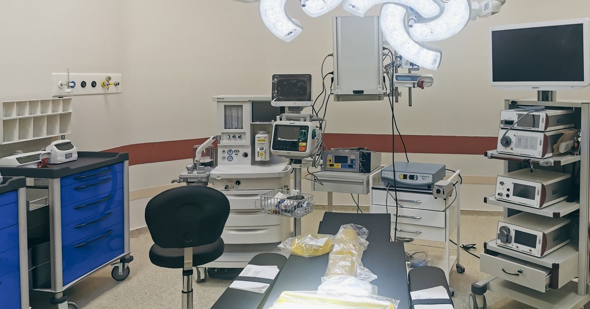
What is necrosectomy?
Necrosectomy means the removal of dead tissue. The word is used most often for the pancreas. In a severe attack of kongosho, part of the gland and the fat around it can lose its blood supply and die. This is called kongosho inayosababisha necrotizing. Over a few weeks the body forms a wall around the dead material, creating a collection known as walled off necrosis.
Other names for necrosectomy
- Necrosectomy ya kongosho
- Pancreatic debridement
- Endoscopic necrosectomy or direct endoscopic necrosectomy
- Video assisted retroperitoneal debridement (VARD)
- Minimally invasive pancreatic necrosectomy
- Open necrosectomy
- Removal of walled off pancreatic necrosis
Why is necrosectomy done?
Most people with pancreatitis recover without any operation. Necrosectomy is considered in a smaller group who have developed necrosis and one of the following problems:
- Infected necrosis: bacteria grow in the dead tissue, causing fever, a rising pulse and worsening blood tests. This is the most common reason and can lead to sepsis if untreated.
- Failure to improve: the person remains unwell for weeks, cannot eat and keeps losing weight even though no infection is proven.
- Kuzuia: the collection presses on the stomach or intestine and causes vomiting, or presses on the bile duct and causes jaundice.
Common causes of the original attack include gongo (Angalia kongosho ya nyongo) and alcohol. Necrosis is different from a simple fluid collection such as a uvimbe bandia wa kongosho, which contains fluid only and is usually easier to drain.
Types of necrosectomy and the step up approach
In the past, early open surgery was the standard treatment. Experience has shown that operating early, while the person is very ill and the dead tissue is not yet separated from healthy tissue, carries a high risk. Current practice follows a step up approach: begin with the least invasive measure that can control the infection and move to the next step only if the person does not improve.
Step 1: Antibiotics and drainage
Antibiotics and supportive care are given first. If a procedure is needed, a thin drainage tube is placed through the skin under CT scan or ultrasound guidance, or a drainage channel is made from the stomach using an endoscope. In a proportion of people, drainage alone is enough and no necrosectomy is needed.
Step 2: Endoscopic necrosectomy
Kutumia ultrasound ya endoscopic, the doctor makes an opening between the stomach or duodenum and the collection and places a stent to keep it open. In later sessions the endoscope is passed through this opening and the dead tissue is pulled out piece by piece. There are no cuts on the skin. This suits collections that lie against the stomach or duodenum.
Step 3: Minimally invasive surgical necrosectomy
If the collection lies to the side or deep in the back of the abdomen, the surgeon can follow the track of a drainage tube through a small cut in the flank and remove the dead tissue under camera vision. This is called video assisted retroperitoneal debridement. Some centres also perform laparoscopic necrosectomy through the stomach.
Step 4: Open necrosectomy
The abdomen is opened through a larger cut, the dead tissue is removed by hand and drains are left in place. Open surgery is now kept for people in whom less invasive methods have failed or are not possible, or when there is another emergency such as bleeding or a hole in the bowel.
| Feature | Endoscopic | Upasuaji usio na uvamizi mdogo | Open |
|---|---|---|---|
| Ufikiaji | Through the mouth and stomach wall | Small cut in the flank or keyhole ports | Large cut on the abdomen |
| Anesthesia | Kutuliza kwa kina au ganzi ya jumla | Anesthesia ya jumla | Anesthesia ya jumla |
| Inafaa kwa | Collections next to the stomach or duodenum | Collections extending to the side or back | Widespread necrosis or failure of other methods |
| Skin wound | hakuna | ndogo | Large, with a risk of later hernia |
Who is suitable for necrosectomy?
The decision is made by a team that includes a surgeon, a gastroenterologist, a radiologist and an intensive care doctor. Necrosectomy is generally considered when:
- Infection of the dead tissue is confirmed or strongly suspected and the person is not improving with antibiotics and drainage.
- The collection has become walled off, which makes removal safer.
- Symptoms such as vomiting, pain or jaundice continue because of the collection.
Nani huenda asifae
- People with sterile necrosis and no symptoms, who are usually watched and not operated on.
- People in the first days or weeks of the illness, in whom the procedure is delayed if their condition allows.
- People with a severe uncorrected bleeding tendency, until it is treated.
How to prepare for necrosectomy
Most people are already in hospital, often in an intensive care or high dependency unit. Preparation is carried out by the treating team.
Uchunguzi
- Vipimo vya damu ikiwa ni pamoja na kuhesabu damu kamili, C protini tendaji, kidney and liver tests, clotting tests and blood sugar. Amylase na lipase will usually have been checked at the start of the illness.
- A contrast enhanced CT scan, and sometimes an MRI, to map the collection, show how much solid material it contains and identify nearby blood vessels.
- An ECG and an anaesthetic assessment.
Dawa za kujadili
- Blood thinners and medicines that affect clotting, which may need to be paused or adjusted.
- Diabetes medicines, as sugar control is often disturbed in pancreatitis.
- Any allergies, particularly to antibiotics, contrast dye or anaesthetic medicines.
Siku hiyo
- You will be asked not to eat or drink for several hours beforehand. If you are being fed through a tube, the feed is stopped for a period advised by the team.
- Fluids and antibiotics are given through a drip.
- The doctor explains the plan, the alternatives and the risks, and you or your family sign a consent form.
How is necrosectomy performed?
The steps differ by method. The outline below describes endoscopic necrosectomy, with notes on the surgical routes.
- Anesthesia: deep sedation or general anaesthesia is given and your pulse, blood pressure and oxygen level are monitored throughout.
- Locating the collection: an endoscope with an ultrasound probe is passed through the mouth into the stomach. The doctor identifies the collection and checks that no large blood vessel lies in the path.
- Creating the opening: a needle and then a wire are passed through the stomach wall into the collection, and a wide metal or plastic stent is placed across the opening. Fluid and pus drain into the stomach.
- Removing dead tissue: in the same or a later session, an endoscope is passed through the stent into the cavity. Loose dead tissue is grasped with snares, baskets or forceps and brought out. The cavity is washed with fluid.
- Vipindi vya kurudia: the procedure is repeated every few days until the cavity is clean. A thin tube may be left through the nose to rinse the cavity between sessions.
- Surgical routes: in video assisted retroperitoneal debridement, a small cut is made in the flank along an existing drain, dead tissue is removed under camera vision and a wash drain is left. In open necrosectomy, the abdomen is opened, the dead tissue is cleared gently by hand and several drains are placed before closure.
Ni nini hufanyika baada ya utaratibu?
You return to the ward or intensive care unit. Pulse, blood pressure, temperature and drain output are checked often. Antibiotics continue, guided by culture results. Nutrition is an important part of recovery; feeding through a tube into the intestine is preferred when eating is not possible, and food by mouth is restarted as soon as it is tolerated. Drains and stents are removed once the collection has resolved, which may take several weeks.
Risks and complications of necrosectomy
People who need this procedure are often seriously ill, so complications are not rare. Some arise from the procedure and some from the disease.
Zaidi ya kawaida
- Bleeding from the cavity wall or nearby blood vessels
- Need for further sessions or a different method
- Ongoing infection or a new jipu
- Leak of pancreatic juice through a drain track (pancreatic fistula)
- Stent blockage or movement
Less common but serious
- A hole in the stomach, bowel or colon, or an abnormal connection between the bowel and the skin
- Heavy bleeding that needs a radiology procedure to block the vessel, or emergency surgery
- Air entering the bloodstream during endoscopic work, which is rare
- Kuganda kwa damu kwenye miguu au mapafu (tazama thrombosis ya mshipa wa kina)
- Failure of other organs, such as the lungs or kidneys, related to severe infection
- A hernia at the wound site after open surgery
- Death, mainly in people who are already critically ill
Longer term effects
- Diabetes, if too little insulin producing tissue remains
- Poor digestion of fat due to lack of pancreatic enzymes (see ukosefu wa kongosho ya exocrine)
- Recurrent collections if the pancreatic duct is disconnected
Results: how well does necrosectomy work?
When infected necrosis is treated in a stepwise way at an experienced centre, most people recover. Studies comparing approaches have shown that starting with drainage and less invasive removal leads to fewer major complications than starting with open surgery. Outcomes depend heavily on how ill the person is, their age, other medical conditions and whether other organs have been affected.
Recovery after necrosectomy
Recovery is gradual. Hospital stay is often measured in weeks, because the underlying illness is severe. After discharge, tiredness, weight loss and weak muscles are common and improve over a few months with good nutrition and gentle activity. People who had open surgery need longer for the wound to heal and should avoid heavy lifting until the surgeon allows it. Follow up visits include blood tests and scans, and a plan to remove any stent or drain.
Wakati wa kutafuta msaada wa matibabu
- Homa au baridi
- Increasing abdominal or back pain
- Kutapika mara kwa mara au kutoweza kuzuia maji mwilini
- Kutapika damu au kutoa kinyesi cheusi
- Njano ya macho au ngozi
- Redness, swelling or discharge at a wound or drain site, or a sudden change in drain output
- Breathlessness, chest pain or swelling of one leg
Life after necrosectomy
- chakula: eat small, frequent meals. A dietitian can advise on protein and energy intake, as utapiamlo is common after a long illness.
- Pombe na tumbaku: avoid alcohol completely and stop smoking, as both raise the risk of further attacks.
- Mawe ya nyongo: if gallstones caused the attack, removal of the gallbladder is usually advised once you have recovered (see cholecystectomy). Some people also need ERCP for stones in the bile duct.
- Sukari ya damu: have your sugar checked at follow up visits, because diabetes can appear months later.
- Digestion: report oily, pale stools or continuing weight loss; enzyme supplements taken with meals may help.
Benefits of necrosectomy
- Removes the source of infection that antibiotics alone cannot clear
- Relieves pressure on the stomach, intestine and bile duct
- Allows eating to restart and nutrition to improve
- Less invasive routes avoid a large wound and reduce stress on the body
- The stepwise approach means many people avoid open surgery
What is the cost of necrosectomy in India?
Gharama inategemea mambo kadhaa:
- The method used (endoscopic, minimally invasive or open) and the number of sessions
- Length of stay in intensive care and in the ward
- Stents, drains and other devices used
- Scans, laboratory tests and antibiotics
- Nutritional support and treatment of complications
- The city, hospital and type of room
Gharama hutofautiana kutoka kwa mtu hadi mtu. Tafadhali wasiliana na hospitali kwa bei halisi.
Most health insurance policies cover medically necessary treatment for pancreatitis and its complications, but check the terms and limits with your insurer.
Why choose Apollo Hospitals for necrosectomy?
- Surgeons, gastroenterologists, radiologists and intensive care doctors plan treatment together.
- Imaging, endoscopy and intensive care services are available within the hospital.
- The step up approach is followed, with the least invasive suitable option considered first.
- Dietitians and physiotherapists support recovery.
- Upatikanaji hutofautiana kati ya vituo. Tafadhali wasiliana na hospitali.
To discuss treatment, you can weka miadi na daktari mkuu wa upasuaji or wasiliana na gastroenterologist katika hospitali za Apollo.
Kusoma kuhusiana
- Kongosho inayosababisha uvimbe
- Pancreatitis
- Vivimbe bandia vya kongosho
- Cholecystectomy ya laparoscopic
- ERCP
Questions to ask your doctor about necrosectomy
- Is the dead tissue infected, and how do you know?
- Can drainage alone be tried first?
- Which method do you recommend for me and why?
- Je, ni vipindi vingapi ninavyoweza kuhitaji?
- How long might I stay in hospital and in intensive care?
- When will the stent or drains be removed?
- Will I need gallbladder surgery later?
- What follow up tests will I need for diabetes and digestion?
Maswali Yanayoulizwa Mara Kwa Mara
1. Is necrosectomy a major procedure?
Yes. Even when it is done through an endoscope without any skin cut, it is performed in people who are seriously ill and it carries real risks. Open necrosectomy is a major abdominal operation.
2. Does everyone with necrotising pancreatitis need necrosectomy?
No. Sterile necrosis without symptoms is usually managed with supportive care and monitoring. A procedure is considered mainly when the dead tissue is infected or causes continuing problems.
3. Why do doctors wait before doing it?
Waiting, when the person's condition allows, lets the dead tissue separate from healthy tissue and become walled off. Removal is then safer, with less bleeding and less damage to living pancreas.
4. What is the step up approach?
It is a staged plan. Treatment begins with antibiotics and drainage. If the person does not improve, dead tissue is removed by an endoscopic or minimally invasive route. Open surgery is used only if these fail or are unsuitable.
5. Is endoscopic necrosectomy better than surgery?
Neither is better for everyone. Endoscopic treatment avoids skin wounds and external leaks of pancreatic juice but often needs several sessions. The choice depends on the position of the collection and the experience of the team.
6. Ni vipindi ngapi vinahitajika?
It varies. Some people need one or two sessions and others need several, a few days apart, until scans and endoscopy show the cavity is clean.
7. Je, nitakuwa macho wakati wa utaratibu?
No. Endoscopic necrosectomy is done under deep sedation or general anaesthesia, and surgical necrosectomy under general anaesthesia.
8. Ahueni huchukua muda gani?
Recovery often takes weeks in hospital and a few months at home before strength and weight return. It depends on the severity of the illness more than on the procedure itself.
9. Can the pancreas work normally afterwards?
Many people keep enough working pancreas. Some develop diabetes or difficulty digesting fat, which can be managed with treatment. Regular follow up helps detect these early.
10. Can necrosis come back?
Dead tissue that has been removed does not return, but new collections can form if the pancreatic duct is damaged or if there is another attack of pancreatitis. Treating the cause, such as gallstones or alcohol use, lowers this risk.
11. What happens to the stent placed in the stomach wall?
It is removed with an endoscope once scans show the collection has resolved.
12. Can I eat during treatment?
Nutrition is encouraged. If you cannot eat, liquid feed is given through a thin tube into the intestine.
13. Is necrosectomy done for other parts of the body?
The term can describe removal of dead tissue anywhere, but in practice it most often refers to the pancreas.
14. Which doctor performs necrosectomy?
Surgical necrosectomy is performed by a gastrointestinal or general surgeon with experience in pancreatic surgery. Endoscopic necrosectomy is performed by an interventional gastroenterologist. Radiologists and intensive care doctors are closely involved.
Hitimisho
Necrosectomy removes dead and infected tissue that can follow severe pancreatitis. Not everyone with necrosis needs it, and when it is required, doctors now favour delayed, stepwise treatment that starts with drainage and uses endoscopic or minimally invasive removal before open surgery. The illness is serious and recovery takes time, but care from an experienced team, good nutrition and treatment of the underlying cause support a good recovery.
Ukurasa huu ni kwa ajili ya taarifa za jumla na hauchukui nafasi ya ushauri kutoka kwa daktari wako. Maamuzi ya matibabu yanapaswa kufanywa na mtaalamu aliyehitimu ambaye anajua historia yako ya matibabu.
Aliongeza hivi karibuni


General and Gastrointestinal Surgeons at Apollo Hospitals










disclaimer:
Taarifa iliyotolewa kwenye ukurasa huu imekusudiwa kwa madhumuni ya jumla ya taarifa na kielimu pekee. Ingawa tunafanya juhudi zinazofaa kuhakikisha kwamba taarifa hiyo ni sahihi, ya kuaminika, na inapitiwa mara kwa mara, haipaswi kuchukuliwa kama mbadala wa ushauri wa kitaalamu wa kimatibabu, utambuzi, au matibabu.
Ufaa wa utaratibu wa kimatibabu, pamoja na faida zake, hatari, maandalizi, kupona, matatizo yanayoweza kutokea, na matokeo yanayotarajiwa, vinaweza kutofautiana kutoka kwa mtu hadi mtu. Mtaalamu wako wa afya ataamua kama utaratibu unafaa kulingana na hali yako binafsi na historia ya matibabu.
Tafadhali wasiliana na mtaalamu wa afya aliyehitimu kwa ushauri wa kibinafsi kabla ya kufanya maamuzi kuhusu utaratibu wowote wa kimatibabu.
Kwa maelezo zaidi kuhusu jinsi maudhui yetu ya kimatibabu yanavyoundwa, kupitiwa, kusasishwa, na kudumishwa, tafadhali soma [Sera yetu ya Uhariri].