


Billede


Neurosciences at Apollo Hospitals, Gurugram, brings together medical and surgical care for conditions affecting the brain, spinal cord, spine, nerves and muscles. Patients may seek care for stroke, seizures, headache, Parkinson's disease, memory problems, nerve or muscle weakness, brain and spine tumors, or injuries involving the head and spine.
Neurological symptoms can have several causes, and some require urgent treatment. A sudden change in speech, facial weakness, loss of movement, a prolonged seizure, severe head injury or new loss of bladder control with limb weakness needs emergency assessment. Neurologists, neurosurgeons, neurointerventionists, critical care specialists, radiologists and rehabilitation professionals may work together according to the diagnosis.
Care can range from clinical assessment and medicines to catheter-based treatment, surgery, critical care and rehabilitation. Imaging and other tests are selected for the individual rather than applied as a standard package. Treatment plans take into account the diagnosis, severity, other medical conditions and the patient's priorities.
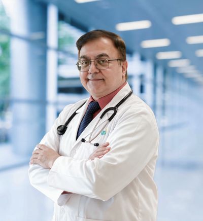
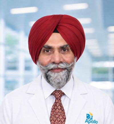
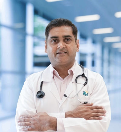
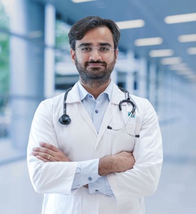
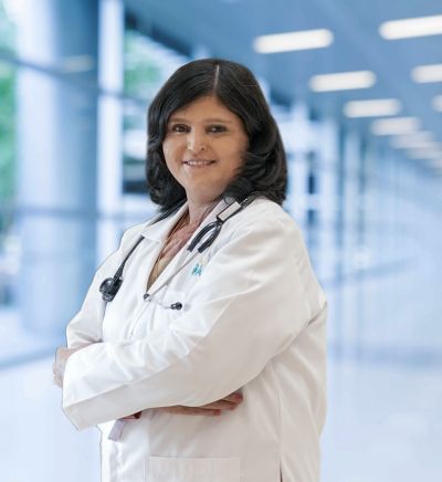
The neurosciences team may include neurologists, neurosurgeons, spine surgeons, neurointerventionists and neurocritical care specialists, supported by neuroradiologists, anesthesiologists, physiotherapists, occupational therapists, speech and swallowing therapists, clinical psychologists and other specialists. Complex cases may be reviewed jointly when medical, catheter-based, surgical and rehabilitation options need to be considered together. Our specialists include:

Eligible patients may receive intravenous thrombolysis within the recommended treatment window. Mechanical thrombectomy may be offered to selected patients with a large-vessel blockage, including some who present later, based on imaging and clinical criteria. Treatment should never be delayed while waiting to see whether symptoms improve.

Treatment may include an antiplatelet medicine or anticoagulant, blood pressure and cholesterol management, diabetes care, smoking cessation and treatment of significant carotid disease. The choice depends on the cause of the stroke and bleeding risk.
Anti-seizure medicine is selected according to seizure type, age, pregnancy potential, other conditions and possible interactions. Doses are adjusted to balance seizure control and side effects, and medicines should not be stopped suddenly without medical advice.
Selected patients with drug-resistant epilepsy may be assessed for removal or disconnection of a seizure-producing area or for a stimulation-based therapy. Evaluation can include prolonged video EEG, specialized imaging and neuropsychological testing.
Management may include medicines for acute attacks, preventive treatment, review of overused pain medicines and lifestyle measures relevant to the individual. Botulinum toxin or newer preventive medicines may be considered for appropriate migraine patterns.
Medicines, physical activity and rehabilitation are adjusted to the person's symptoms and function. Botulinum toxin may be used for selected dystonia or spasticity. Deep brain stimulation may be considered for carefully selected patients with Parkinson's disease, tremor or dystonia.
Steroids, intravenous immunoglobulin, plasma exchange or other immune-modifying medicines may be used for selected conditions such as Guillain-Barré syndrome, myasthenia gravis, multiple sclerosis or autoimmune encephalitis. Choice and timing depend on the diagnosis and severity.
Surgery may be used to obtain a diagnosis, remove as much tumor as can be done safely, relieve pressure or stabilize the spine. The approach depends on tumor type, location and the functions at risk.
Some pituitary tumors respond to medicine. Others may require endoscopic surgery through the nose, radiotherapy or a combination of treatments, with endocrinology and ophthalmology input when needed.
Options may include observation, catheter-based treatment such as coiling or flow diversion, or surgical clipping. Anatomy, rupture status, age and overall health influence the recommendation.
Procedures may include discectomy, decompression, fusion or stabilization. Minimally invasive or endoscopic approaches may be suitable for selected patients, but the safest effective approach is chosen after clinical and imaging assessment.
Intensive monitoring and organ support may be required for severe stroke, brain injury, status epilepticus, infection or after major neurosurgery.
Therapy after stroke, injury or surgery may address mobility, self-care, speech, swallowing, cognition and return to daily activities. Rehabilitation should begin when the patient is medically stable and continue according to goals and progress.
Rapid CT, CT angiography, MRI or perfusion imaging can help identify the type of stroke, the vessel involved and whether tissue may still be salvageable. The exact pathway depends on presentation and local capability.
Angiography equipment provides live imaging during catheter-based diagnosis and treatment of selected strokes, aneurysms and vascular malformations.
Navigation systems relate surgical instruments to the patient's preoperative scan and can help the surgeon plan and follow a route to the target. They support surgical precision but do not remove the risks of brain or spine surgery.
Electrical signals from the brain, spinal cord, nerves or muscles can be monitored during selected operations to alert the team to changes in neurological function.
Magnification, illumination and endoscopic visualization can support surgery through smaller or more direct corridors in selected brain, pituitary and spine procedures.
Synchronized video and EEG can record events over an extended period and support diagnosis, medication adjustment and evaluation for epilepsy surgery.
Selected patients may need continuous EEG, intracranial pressure monitoring or other bedside neurological monitoring in intensive care.
Mobility aids, swallowing assessment tools and task-specific therapy equipment may support recovery and safety during rehabilitation.


Sudden facial drooping, weakness or numbness on one side, difficulty speaking, loss of balance or sudden visual loss may indicate stroke. Note when the person was last known to be well, call 112 and go to an emergency department immediately. Do not wait for the symptoms to settle.
Possibly. Intravenous clot-dissolving treatment is generally considered within 4.5 hours for eligible patients, and selected patients may qualify beyond standard windows based on advanced imaging. Mechanical thrombectomy can benefit some large-vessel strokes for up to 24 hours after the person was last known to be well. Earlier arrival still provides the widest range of options.
A neurologist diagnoses and treats nervous-system conditions mainly through medicines and non-surgical care. A neurosurgeon performs operations involving the brain, spine and nerves. They may assess a patient together when the best approach is uncertain or several treatments are possible.
No. Primary headache disorders such as migraine and tension-type headache are much more common. Urgent assessment is needed for a sudden severe headache, headache with fever and neck stiffness, new weakness or confusion, seizure, visual loss or a rapidly changing pattern.
MRI is painless and does not use ionizing radiation, but the scanner is noisy and enclosed. Every pacemaker, implant, metal fragment or device must be checked for MRI safety. Some are MRI conditional and require specific precautions. Sedation may be considered only after appropriate assessment and monitoring arrangements.
No. A routine EEG records brain activity only during the test, so it may be normal between seizures. The history remains central, and repeat, sleep-deprived or prolonged video EEG may be useful when the diagnosis is uncertain.
Move dangerous objects away, place something soft under the head and time the seizure. Do not restrain the person or put anything in the mouth. Turn them onto their side when the movements stop and stay with them. Call emergency services if the seizure lasts five minutes, another begins without recovery, breathing is difficult, injury occurs or it is the first known seizure.
Many people achieve seizure freedom with appropriate treatment, but some continue to have seizures despite medicine. If two suitable anti-seizure medicine schedules have failed, referral to an epilepsy specialist is appropriate because surgery or another therapy may be considered.
Many people with epilepsy work and have healthy pregnancies. Safety-sensitive work and driving depend on seizure control and applicable legal requirements. Pregnancy should be planned with the neurologist because medicine choice and dose may need review before conception. Anti-seizure medicine should never be stopped without medical advice.
There is currently no cure, but medicines, physical activity and therapy can improve symptoms and function. Deep brain stimulation may help selected patients whose symptoms or medication-related fluctuations are difficult to control, but it does not stop the disease from progressing.
No. Many cases improve with activity modification, pain management and physiotherapy. Surgical assessment becomes more important when pain remains severe despite treatment or when there is progressive weakness. New bladder or bowel difficulty, numbness around the groin or rapidly worsening weakness requires emergency care.
No. Brain tumors include benign and malignant types, and cancer from another organ can also spread to the brain. Imaging provides important clues, but tissue examination is often needed to identify the exact type and guide treatment.
No. The surgical goal depends on the tumor type, location and the neurological functions nearby. Sometimes complete removal can be attempted safely. In other cases, biopsy, partial removal or non-surgical treatment may offer a better balance of benefit and risk.
The risk depends on the area of the brain involved and the planned procedure. Imaging, navigation, monitoring and awake mapping may help protect important functions in selected cases, but they cannot remove all risk. The surgeon should explain the individual risks before consent.
Recovery varies widely with the size and location of the stroke, complications, previous health and access to rehabilitation. Improvement is often fastest in the first weeks and months but may continue for much longer. Therapy goals are reviewed as the person progresses.
No. Sleep problems, depression, hearing loss, thyroid disease, vitamin deficiency, medication effects, infection and other conditions can affect memory. Assessment combines the history, cognitive testing and selected investigations to identify treatable contributors and determine whether a neurodegenerative condition is present.



 Bedste hospital i nærheden af mig, Chennai
Bedste hospital i nærheden af mig, Bangalore
Bedste hospital i nærheden af mig Hyderabad
Bedste hospital i nærheden af mig, Mumbai
Bedste hospital i nærheden af mig Delhi
Bedste hospital i nærheden af mig, Kolkata
Bedste hospital i nærheden af mig i Pune
Bedste hospital i nærheden af mig, Ahmedabad
Bedste hospital i nærheden af mig Bhubaneswar
Bedste hospital i nærheden af mig i Bilaspur
Bedste hospital i nærheden af mig, Guwahati
Bedste hospital i nærheden af mig i Indore
Bedste hospital i nærheden af mig i Kochi
Bedste hospital i nærheden af mig, Lucknow
Bedste hospital i nærheden af mig Madurai
Bedste hospital i nærheden af mig i Mysore
Bedste hospital i nærheden af mig i Nashik
Bedste hospital i nærheden af mig i Noida
Bedste hospital i nærheden af mig Rourkela
Bedste hospital i nærheden af mig Visakhapatnam
Kardiolog i nærheden af mig i Chennai
Kardiolog i nærheden af mig i Bangalore
Kardiolog i nærheden af mig i Hyderabad
Kardiolog i nærheden af mig i Mumbai
Kardiolog i nærheden af mig i Delhi
Kardiolog i nærheden af mig i Kolkata
Kardiolog i nærheden af mig i Pune
Kardiolog i nærheden af mig i Ahmedabad
Kardiolog i nærheden af mig i Bhubaneswar
Kardiolog i nærheden af mig i Bilaspur
Kardiolog i nærheden af mig i Guwahati
Kardiolog i nærheden af mig i Indore
Kardiolog i nærheden af mig i Kochi
Kardiolog i nærheden af mig i Lucknow
Kardiolog i nærheden af mig i Madurai
Kardiolog i nærheden af mig i Mysore
Kardiolog i nærheden af mig i Nashik
Kardiolog i nærheden af mig i Noida
Kardiolog i nærheden af mig i Rourkela
Kardiolog i nærheden af mig i Visakhapatnam
Neurolog i nærheden af mig i Chennai
Neurolog i nærheden af mig i Bangalore
Neurolog i nærheden af mig i Hyderabad
Neurolog i nærheden af mig i Mumbai
Neurolog i nærheden af mig i Delhi
Neurolog i nærheden af mig i Kolkata
Neurolog i nærheden af mig i Ahmedabad
Neurolog i nærheden af mig i Bhubaneswar
Neurolog i nærheden af mig i Bilaspur
Neurolog i nærheden af mig i Guwahati
Neurolog i nærheden af mig i Indore
Neurolog i nærheden af mig i Kochi
Neurolog i nærheden af mig i Lucknow
Neurolog i nærheden af mig i Madurai
Neurolog i nærheden af mig i Mysore
Neurolog i nærheden af mig i Nashik
Neurolog i nærheden af mig i Noida
Neurolog i nærheden af mig i Rourkela
Neurolog i nærheden af mig i Visakhapatnam
Ortopædkirurg i nærheden af mig i Chennai
Ortopædlæge i nærheden af mig i Bangalore
Ortopædkirurg i nærheden af mig i Hyderabad
Ortopædlæge i nærheden af mig i Mumbai
Ortopædlæge i nærheden af mig i Delhi
Ortopædlæge i nærheden af mig i Kolkata
Ortopædlæge i nærheden af mig i Pune
Ortopædkirurg i nærheden af mig i Ahmedabad
Ortopædlæge i nærheden af mig i Bhubaneswar
Ortopædkirurg i nærheden af mig i Bilaspur
Ortopædlæge i nærheden af mig i Guwahati
Ortopædlæge i nærheden af mig i Indore
Ortopædlæge i nærheden af mig i Kochi
Ortopædkirurg i nærheden af mig i Lucknow
Ortopædlæge i nærheden af mig i Madurai
Ortopædlæge i nærheden af mig i Mysore
Ortopædlæge i nærheden af mig i Nashik
Ortopædlæge i nærheden af mig i Noida
Ortopæder i nærheden af mig i Rourkela
Ortopædkirurg i nærheden af mig i Visakhapatnam
Onkolog i nærheden af mig i Chennai
Onkolog i nærheden af mig i Bangalore
Onkolog i nærheden af mig i Hyderabad
Onkolog i nærheden af mig i Mumbai
Onkolog i nærheden af mig i Delhi
Onkolog i nærheden af mig i Kolkata
Onkolog i nærheden af mig i Pune
Onkolog i nærheden af mig i Ahmedabad
Onkolog i nærheden af mig i Bhubaneswar
Onkolog i nærheden af mig i Bilaspur
Onkolog i nærheden af mig i Guwahati
Onkolog i nærheden af mig i Indore
Onkolog i nærheden af mig i Kochi
Onkolog i nærheden af mig i Lucknow
Onkolog i nærheden af mig i Madurai
Onkolog i nærheden af mig i Mysore
Onkolog i nærheden af mig i Nashik
Onkolog i nærheden af mig i Noida
Onkolog i nærheden af mig i Rourkela
Gastroenterolog i nærheden af mig i Chennai
Gastroenterolog i nærheden af mig i Bangalore
Gastroenterolog i nærheden af mig i Hyderabad
Gastroenterolog i nærheden af mig i Mumbai
Gastroenterolog i nærheden af mig i Delhi
Gastroenterolog i nærheden af mig i Kolkata
Gastroenterolog i nærheden af mig i Pune
Gastroenterolog i nærheden af mig i Ahmedabad
Gastroenterolog i nærheden af mig i Bhubaneswar
Gastroenterolog i nærheden af mig i Bilaspur
Gastroenterolog i nærheden af mig i Guwahati
Gastroenterolog i nærheden af mig i Indore
Gastroenterolog i nærheden af mig i Kochi
Gastroenterolog i nærheden af mig i Lucknow
Gastroenterolog i nærheden af mig i Madurai
Gastroenterolog i nærheden af mig i Mysore
Gastroenterolog i nærheden af mig i Nashik
Gastroenterolog i nærheden af mig i Noida
Gastroenterolog i nærheden af mig i Rourkela
Gastroenterolog i nærheden af mig i Visakhapatnam
Transplantationskirurg i nærheden af mig i Chennai
Transplantationskirurg i nærheden af mig i Bangalore
Transplantationskirurg i nærheden af mig i Hyderabad
Transplantationskirurg i nærheden af mig i Mumbai
Transplantationskirurg i nærheden af mig i Delhi
Transplantationskirurg i nærheden af mig i Kolkata
Transplantationskirurg i nærheden af mig i Indore
Transplantationskirurg i nærheden af mig i Kochi
Transplantationskirurg i nærheden af mig i Lucknow
Transplantationskirurg i nærheden af mig i Mysore
ØNH-specialist i nærheden af mig i Chennai
ØNH-specialist i nærheden af mig i Bangalore
ØNH-specialist i nærheden af mig i Hyderabad
ØNH-specialist i nærheden af mig i Mumbai
ØNH-specialist i nærheden af mig i Delhi
ØNH-specialist i nærheden af mig i Kolkata
ØNH-specialist i nærheden af mig i Pune
ØNH-specialist i nærheden af mig i Ahmedabad
ØNH-specialist i nærheden af mig i Bhubaneswar
ØNH-specialist i nærheden af mig i Bilaspur
ØNH-specialist i nærheden af mig i Guwahati
ØNH-specialist i nærheden af mig i Indore
ØNH-specialist i nærheden af mig i Kochi
ØNH-specialist i nærheden af mig i Lucknow
ØNH-specialist i nærheden af mig i Madurai
ØNH-specialist i nærheden af mig i Mysore
ØNH-specialist i nærheden af mig i Nashik
ØNH-specialist i nærheden af mig i Noida
ØNH-specialist i nærheden af mig i Rourkela
ØNH-specialist i nærheden af mig i Visakhapatnam
Lungelæge i nærheden af mig i Chennai
Lungelæge i nærheden af mig i Bangalore
Lungelæge i nærheden af mig i Hyderabad
Lungelæge i nærheden af mig i Mumbai
Lungelæge i nærheden af mig i Delhi
Lungelæge i nærheden af mig i Kolkata
Lungelæge i nærheden af mig i Ahmedabad
Lungelæge i nærheden af mig i Bhubaneswar
Lungelæge i nærheden af mig i Bilaspur
Lungelæge i nærheden af mig i Guwahati
Lungelæge i nærheden af mig i Indore
Lungelæge i nærheden af mig i Kochi
Lungelæge i nærheden af mig i Lucknow
Lungelæge i nærheden af mig i Madurai
Lungelæge i nærheden af mig i Mysore
Lungelæge i nærheden af mig i Nashik
Lungelæge i nærheden af mig i Noida
Lungelæge i nærheden af mig i Rourkela
Lungelæge i nærheden af mig i Visakhapatnam
Tandlæge i nærheden af mig i Chennai
Tandlæge i nærheden af mig i Bangalore
Tandlæge i nærheden af mig i Hyderabad
Tandlæge i nærheden af mig i Delhi
Tandlæge i nærheden af mig i Kolkata
Tandlæge i nærheden af mig i Ahmedabad
Tandlæge i nærheden af mig i Bhubaneswar
Tandlæge i nærheden af mig i Bilaspur
Tandlæge i nærheden af mig i Guwahati
Tandlæge i nærheden af mig i Kochi
Tandlæge i nærheden af mig i Lucknow
Tandlæge i nærheden af mig i Madurai
Tandlæge i nærheden af mig i Nashik
Tandlæge i nærheden af mig i Noida
Tandlæge i nærheden af mig i Rourkela
Tandlæge i nærheden af mig i Visakhapatnam
Børnelæge i nærheden af mig i Chennai
Børnelæge i nærheden af mig i Bangalore
Børnelæge i nærheden af mig i Hyderabad
Børnelæge i nærheden af mig i Mumbai
Børnelæge i nærheden af mig i Delhi
Børnelæge i nærheden af mig i Kolkata
Børnelæge i nærheden af mig i Pune
Børnelæge i nærheden af mig i Ahmedabad
Børnelæge i nærheden af mig i Bhubaneswar
Børnelæge i nærheden af mig i Bilaspur
Børnelæge i nærheden af mig i Guwahati
Børnelæge i nærheden af mig i Indore
Børnelæge i nærheden af mig i Kochi
Børnelæge i nærheden af mig i Lucknow
Børnelæge i nærheden af mig i Madurai
Børnelæge i nærheden af mig i Mysore
Børnelæge i nærheden af mig i Nashik
Børnelæge i nærheden af mig i Noida
Børnelæge i nærheden af mig i Visakhapatnam
Hudlæge i nærheden af mig i Chennai
Hudlæge i nærheden af mig i Bangalore
Hudlæge i nærheden af mig i Hyderabad
Hudlæge i nærheden af mig i Delhi
Hudlæge i nærheden af mig i Kolkata
Hudlæge i nærheden af mig i Ahmedabad
Hudlæge i nærheden af mig i Bhubaneswar
Hudlæge i nærheden af mig i Bilaspur
Hudlæge i nærheden af mig i Guwahati
Hudlæge i nærheden af mig i Indore
Hudlæge i nærheden af mig i Kochi
Hudlæge i nærheden af mig i Lucknow
Hudlæge i nærheden af mig i Madurai
Hudlæge i nærheden af mig i Mysore
Hudlæge i nærheden af mig i Noida
Hudlæge i nærheden af mig i Visakhapatnam
Urolog i nærheden af mig i Chennai
Urolog i nærheden af mig i Bangalore
Urolog i nærheden af mig i Hyderabad
Urolog i nærheden af mig i Delhi
Urolog i nærheden af mig i Kolkata
Urolog i nærheden af mig i Pune
Urolog i nærheden af mig i Ahmedabad
Urolog i nærheden af mig i Bhubaneswar
Urolog i nærheden af mig i Bilaspur
Urolog i nærheden af mig i Guwahati
Urolog i nærheden af mig i Indore
Urolog i nærheden af mig i Kochi
Urolog i nærheden af mig i Lucknow
Urolog i nærheden af mig i Madurai
Urolog i nærheden af mig i Mysore
Urolog i nærheden af mig i Nashik
Urolog i nærheden af mig i Noida
Urolog i nærheden af mig i Rourkela
Urolog i nærheden af mig i Visakhapatnam
Endokrinolog og diabetolog i nærheden af mig i Chennai
Endokrinolog og diabetolog i nærheden af mig i Bangalore
Endokrinolog og diabetolog i nærheden af mig i Hyderabad
Endokrinolog og diabetolog i nærheden af mig i Mumbai
Endokrinolog og diabetolog i nærheden af mig i Delhi
Endokrinolog og diabetolog i nærheden af mig i Kolkata
Endokrinolog og diabetolog i nærheden af mig i Pune
Endokrinolog og diabetolog i nærheden af mig i Ahmedabad
Endokrinolog og diabetolog i nærheden af mig i Bhubaneswar
Endokrinolog og diabetolog i nærheden af mig i Bilaspur
Endokrinolog og diabetolog i nærheden af mig i Guwahati
Endokrinolog og diabetolog i nærheden af mig i Indore
Endokrinolog og diabetolog i nærheden af mig i Kochi
Endokrinolog og diabetolog i nærheden af mig i Lucknow
Endokrinolog og diabetolog i nærheden af mig i Madurai
Endokrinolog og diabetolog i nærheden af mig i Mysore
Endokrinolog og diabetolog i nærheden af mig i Noida
Endokrinolog og diabetolog i nærheden af mig i Rourkela
Endokrinolog og diabetolog i nærheden af mig i Visakhapatnam
Gynækolog i nærheden af mig i Chennai
Gynækolog i nærheden af mig i Bangalore
Gynækolog i nærheden af mig i Hyderabad
Gynækolog i nærheden af mig i Mumbai
Gynækolog i nærheden af mig i Delhi
Gynækolog i nærheden af mig i Kolkata
Gynækolog i nærheden af mig i Ahmedabad
Gynækolog i nærheden af mig i Bhubaneswar
Gynækolog i nærheden af mig i Bilaspur
Gynækolog i nærheden af mig i Guwahati
Gynækolog i nærheden af mig i Indore
Gynækolog i nærheden af mig i Kochi
Gynækolog i nærheden af mig i Lucknow
Gynækolog i nærheden af mig i Madurai
Gynækolog i nærheden af mig i Mysore
Gynækolog i nærheden af mig i Nashik
Gynækolog i nærheden af mig i Noida
Gynækolog i nærheden af mig i Rourkela
Gynækolog i nærheden af mig i Visakhapatnam
Praktiserende læge i nærheden af mig i Chennai
Praktiserende læge i nærheden af mig i Bangalore
Praktiserende læge i nærheden af mig i Hyderabad
Praktiserende læge i nærheden af mig i Mumbai
Praktiserende læge i nærheden af mig i Delhi
Praktiserende læge i nærheden af mig i Kolkata
Praktiserende læge i nærheden af mig i Pune
Praktiserende læge i nærheden af mig i Ahmedabad
Praktiserende læge i nærheden af mig i Bhubaneswar
Praktiserende læge i nærheden af mig i Bilaspur
Praktiserende læge i nærheden af mig i Guwahati
Praktiserende læge i nærheden af mig i Indore
Praktiserende læge i nærheden af mig i Kochi
Praktiserende læge i nærheden af mig i Lucknow
Praktiserende læge i nærheden af mig i Madurai
Praktiserende læge i nærheden af mig i Mysore
Praktiserende læge i nærheden af mig i Nashik
Praktiserende læge i nærheden af mig i Noida
Praktiserende læge i nærheden af mig i Visakhapatnam
Psykolog i nærheden af mig i Chennai
Psykolog i nærheden af mig i Bangalore
Psykolog i nærheden af mig i Hyderabad
Psykolog i nærheden af mig i Mumbai
Psykolog i nærheden af mig i Delhi
Psykolog i nærheden af mig i Kolkata
Psykolog i nærheden af mig i Ahmedabad
Psykolog i nærheden af mig i Bhubaneswar
Psykolog i nærheden af mig i Bilaspur
Psykolog i nærheden af mig i Guwahati
Psykolog i nærheden af mig i Indore
Psykolog i nærheden af mig i Kochi
Psykolog i nærheden af mig i Lucknow
Psykolog i nærheden af mig i Madurai
Psykolog i nærheden af mig i Mysore
Psykolog i nærheden af mig i Nashik
Psykolog i nærheden af mig i Noida
Psykolog i nærheden af mig i Visakhapatnam
Almen kirurg i nærheden af mig i Chennai
Almen kirurg i nærheden af mig i Bangalore
Almen kirurg i nærheden af mig i Hyderabad
Almen kirurg i nærheden af mig i Mumbai
Almen kirurg i nærheden af mig i Delhi
Almen kirurg i nærheden af mig i Kolkata
Almen kirurg i nærheden af mig i Pune
Almen kirurg i nærheden af mig i Ahmedabad
Almen kirurg i nærheden af mig i Bhubaneswar
Almen kirurg i nærheden af mig i Bilaspur
Almen kirurg i nærheden af mig i Guwahati
Almen kirurg i nærheden af mig i Indore
Almen kirurg i nærheden af mig i Lucknow
Almen kirurg i nærheden af mig i Madurai
Almen kirurg i nærheden af mig i Mysore
Almen kirurg i nærheden af mig i Nashik
Almen kirurg i nærheden af mig i Noida
Almen kirurg i nærheden af mig i Rourkela
Bedste hospital i Karukutty, Cochin
Bedste hospital i Greams Road, Chennai
Bedste hospital i Kuvempunagar, Mysore
Bedste hospital i Vanagaram, Chennai
Bedste hospital i Teynampet, Chennai
Bedste hospital i OMR, Chennai
Bedste kræfthospital i Bhat, Gandhinagar, Ahmedabad
Bedste kræfthospital i Electronic City, Bangalore
Bedste kræfthospital i Teynampet, Chennai
Bedste kræfthospital i HSR Layout, Bangalore
Bedste Protonkræftcenter i Chennai
Bedste børnehospital i Thousand Lights, Chennai
Bedste kvindehospital i Thousand Lights, Chennai
Bedste hospital i Paschim Boragaon, Guwahati
Bedste hospital i PH Road, Chennai
Bedste hjertecenter i Thousand Lights, Chennai
Bedste hospital i Jubilee Hills, Hyderabad
Bedste hospital i Tondiarpet, Chennai
Bedste hospital i Kotturpuram, Chennai
Bedste hospital i Kovai Road, Karur
Bedste hospital i Karapakkam, Chennai
Bedste hospital i Arilova, Vizag
Bedste hospital i Kanpur Road, Lucknow
Bedste hospital i sektor 26, Noida
Bedste hospital i Gandhinagar, Ahmedabad
Bedste hospital i Aragonda, Andhra Pradesh
Bedste hospital i Bannerghatta Road, Bangalore
Bedste hospital i Unit-15, Bhubaneswar
Bedste hospital i Seepat Road, Bilaspur
Bedste hospital i Ellisbridge, Ahmedabad
Det bedste hospital i New Delhi
Bedste hospital i DRDO, Hyderabad
Bedste hospital i GS Road, Guwahati
Bedste hospital i Hyderguda, Hyderabad
Bedste hospital i Vijay Nagar, Indore
Bedste hospital i Suryaraopeta Main Road, Kakinada
Bedste hospital i Canal Circular Road, Kolkata
Bedste hospital i CBD Belapur, Navi Mumbai
Bedste hospital i Panchavati, Nashik
Bedste hospital i Secunderabad, Hyderabad
Bedste hospital i Seshadripuram, Bangalore
Bedste hospital i Waltair Main Road, Visakhapatnam
Bedste hospital i Subhash Nagar Road, Karimnagar
Bedste hospital i Managari, Karaikudi
Bedste hospital i Arepally, Warangal
Bedste hospital i Arera Colony, Bhopal
Bedste hospital i Jayanagar, Bangalore
Bedste hospital i KK Nagar, Madurai
Bedste hospital i Ramji Nagar, Nellore
Bedste hospital i sektor-19, Rourkela
Bedste hospital i Swargate, Pune
Det bedste hospital for kvinders kræft i det sydlige Delhi
CABG
CAR T celleterapi
Laparoskopisk cholecystektomi
hysterektomi
Nyretransplantation
Ekstrakorporal Shockwave Lithotripsi
Levertransplantation
Lung Transplantation
Hip Arthroscopy
Total hofteudskiftning
Protonterapi
Minimalt invasiv Subvastus total knæudskiftning
Fast Track Daycare knæalloplastik
Sleeve Gastrectomy
Lasik kirurgi
Rhinoplasty
Fedtsugning
Koronar angiogram
Transkateter aorta ventil udskiftning
MitraClip-ventilreparation
Minimalt invasiv hjertekirurgi
Kateter ablation
ACL genopbygningskirurgi
Omvendt skulderudskiftning
Endometrial Ablation
Embolisering af livmoderarterie
Ovariecystektomi
Brystkræftkirurgi
Brachyterapi
koloskopi
polypektomi
Deep Brain Stimulation
Peritoneal dialyse
Nyrebiopsi
Parathyreoidektomi
Cytoreduktiv kirurgi
Keramisk total knæalloplastik
ERCP
Bedste hospital i nærheden af mig, Chennai
Bedste hospital i nærheden af mig, Bangalore
Bedste hospital i nærheden af mig Hyderabad
Bedste hospital i nærheden af mig, Mumbai
Bedste hospital i nærheden af mig Delhi
Bedste hospital i nærheden af mig, Kolkata
Bedste hospital i nærheden af mig i Pune
Bedste hospital i nærheden af mig, Ahmedabad
Bedste hospital i nærheden af mig Bhubaneswar
Bedste hospital i nærheden af mig i Bilaspur
Bedste hospital i nærheden af mig, Guwahati
Bedste hospital i nærheden af mig i Indore
Bedste hospital i nærheden af mig i Kochi
Bedste hospital i nærheden af mig, Lucknow
Bedste hospital i nærheden af mig Madurai
Bedste hospital i nærheden af mig i Mysore
Bedste hospital i nærheden af mig i Nashik
Bedste hospital i nærheden af mig i Noida
Bedste hospital i nærheden af mig Rourkela
Bedste hospital i nærheden af mig Visakhapatnam
Kardiolog i nærheden af mig i Chennai
Kardiolog i nærheden af mig i Bangalore
Kardiolog i nærheden af mig i Hyderabad
Kardiolog i nærheden af mig i Mumbai
Kardiolog i nærheden af mig i Delhi
Kardiolog i nærheden af mig i Kolkata
Kardiolog i nærheden af mig i Pune
Kardiolog i nærheden af mig i Ahmedabad
Kardiolog i nærheden af mig i Bhubaneswar
Kardiolog i nærheden af mig i Bilaspur
Kardiolog i nærheden af mig i Guwahati
Kardiolog i nærheden af mig i Indore
Kardiolog i nærheden af mig i Kochi
Kardiolog i nærheden af mig i Lucknow
Kardiolog i nærheden af mig i Madurai
Kardiolog i nærheden af mig i Mysore
Kardiolog i nærheden af mig i Nashik
Kardiolog i nærheden af mig i Noida
Kardiolog i nærheden af mig i Rourkela
Kardiolog i nærheden af mig i Visakhapatnam
Neurolog i nærheden af mig i Chennai
Neurolog i nærheden af mig i Bangalore
Neurolog i nærheden af mig i Hyderabad
Neurolog i nærheden af mig i Mumbai
Neurolog i nærheden af mig i Delhi
Neurolog i nærheden af mig i Kolkata
Neurolog i nærheden af mig i Ahmedabad
Neurolog i nærheden af mig i Bhubaneswar
Neurolog i nærheden af mig i Bilaspur
Neurolog i nærheden af mig i Guwahati
Neurolog i nærheden af mig i Indore
Neurolog i nærheden af mig i Kochi
Neurolog i nærheden af mig i Lucknow
Neurolog i nærheden af mig i Madurai
Neurolog i nærheden af mig i Mysore
Neurolog i nærheden af mig i Nashik
Neurolog i nærheden af mig i Noida
Neurolog i nærheden af mig i Rourkela
Neurolog i nærheden af mig i Visakhapatnam
Ortopædkirurg i nærheden af mig i Chennai
Ortopædlæge i nærheden af mig i Bangalore
Ortopædkirurg i nærheden af mig i Hyderabad
Ortopædlæge i nærheden af mig i Mumbai
Ortopædlæge i nærheden af mig i Delhi
Ortopædlæge i nærheden af mig i Kolkata
Ortopædlæge i nærheden af mig i Pune
Ortopædkirurg i nærheden af mig i Ahmedabad
Ortopædlæge i nærheden af mig i Bhubaneswar
Ortopædkirurg i nærheden af mig i Bilaspur
Ortopædlæge i nærheden af mig i Guwahati
Ortopædlæge i nærheden af mig i Indore
Ortopædlæge i nærheden af mig i Kochi
Ortopædkirurg i nærheden af mig i Lucknow
Ortopædlæge i nærheden af mig i Madurai
Ortopædlæge i nærheden af mig i Mysore
Ortopædlæge i nærheden af mig i Nashik
Ortopædlæge i nærheden af mig i Noida
Ortopæder i nærheden af mig i Rourkela
Ortopædkirurg i nærheden af mig i Visakhapatnam
Onkolog i nærheden af mig i Chennai
Onkolog i nærheden af mig i Bangalore
Onkolog i nærheden af mig i Hyderabad
Onkolog i nærheden af mig i Mumbai
Onkolog i nærheden af mig i Delhi
Onkolog i nærheden af mig i Kolkata
Onkolog i nærheden af mig i Pune
Onkolog i nærheden af mig i Ahmedabad
Onkolog i nærheden af mig i Bhubaneswar
Onkolog i nærheden af mig i Bilaspur
Onkolog i nærheden af mig i Guwahati
Onkolog i nærheden af mig i Indore
Onkolog i nærheden af mig i Kochi
Onkolog i nærheden af mig i Lucknow
Onkolog i nærheden af mig i Madurai
Onkolog i nærheden af mig i Mysore
Onkolog i nærheden af mig i Nashik
Onkolog i nærheden af mig i Noida
Onkolog i nærheden af mig i Rourkela
Gastroenterolog i nærheden af mig i Chennai
Gastroenterolog i nærheden af mig i Bangalore
Gastroenterolog i nærheden af mig i Hyderabad
Gastroenterolog i nærheden af mig i Mumbai
Gastroenterolog i nærheden af mig i Delhi
Gastroenterolog i nærheden af mig i Kolkata
Gastroenterolog i nærheden af mig i Pune
Gastroenterolog i nærheden af mig i Ahmedabad
Gastroenterolog i nærheden af mig i Bhubaneswar
Gastroenterolog i nærheden af mig i Bilaspur
Gastroenterolog i nærheden af mig i Guwahati
Gastroenterolog i nærheden af mig i Indore
Gastroenterolog i nærheden af mig i Kochi
Gastroenterolog i nærheden af mig i Lucknow
Gastroenterolog i nærheden af mig i Madurai
Gastroenterolog i nærheden af mig i Mysore
Gastroenterolog i nærheden af mig i Nashik
Gastroenterolog i nærheden af mig i Noida
Gastroenterolog i nærheden af mig i Rourkela
Gastroenterolog i nærheden af mig i Visakhapatnam
Transplantationskirurg i nærheden af mig i Chennai
Transplantationskirurg i nærheden af mig i Bangalore
Transplantationskirurg i nærheden af mig i Hyderabad
Transplantationskirurg i nærheden af mig i Mumbai
Transplantationskirurg i nærheden af mig i Delhi
Transplantationskirurg i nærheden af mig i Kolkata
Transplantationskirurg i nærheden af mig i Indore
Transplantationskirurg i nærheden af mig i Kochi
Transplantationskirurg i nærheden af mig i Lucknow
Transplantationskirurg i nærheden af mig i Mysore
ØNH-specialist i nærheden af mig i Chennai
ØNH-specialist i nærheden af mig i Bangalore
ØNH-specialist i nærheden af mig i Hyderabad
ØNH-specialist i nærheden af mig i Mumbai
ØNH-specialist i nærheden af mig i Delhi
ØNH-specialist i nærheden af mig i Kolkata
ØNH-specialist i nærheden af mig i Pune
ØNH-specialist i nærheden af mig i Ahmedabad
ØNH-specialist i nærheden af mig i Bhubaneswar
ØNH-specialist i nærheden af mig i Bilaspur
ØNH-specialist i nærheden af mig i Guwahati
ØNH-specialist i nærheden af mig i Indore
ØNH-specialist i nærheden af mig i Kochi
ØNH-specialist i nærheden af mig i Lucknow
ØNH-specialist i nærheden af mig i Madurai
ØNH-specialist i nærheden af mig i Mysore
ØNH-specialist i nærheden af mig i Nashik
ØNH-specialist i nærheden af mig i Noida
ØNH-specialist i nærheden af mig i Rourkela
ØNH-specialist i nærheden af mig i Visakhapatnam
Lungelæge i nærheden af mig i Chennai
Lungelæge i nærheden af mig i Bangalore
Lungelæge i nærheden af mig i Hyderabad
Lungelæge i nærheden af mig i Mumbai
Lungelæge i nærheden af mig i Delhi
Lungelæge i nærheden af mig i Kolkata
Lungelæge i nærheden af mig i Ahmedabad
Lungelæge i nærheden af mig i Bhubaneswar
Lungelæge i nærheden af mig i Bilaspur
Lungelæge i nærheden af mig i Guwahati
Lungelæge i nærheden af mig i Indore
Lungelæge i nærheden af mig i Kochi
Lungelæge i nærheden af mig i Lucknow
Lungelæge i nærheden af mig i Madurai
Lungelæge i nærheden af mig i Mysore
Lungelæge i nærheden af mig i Nashik
Lungelæge i nærheden af mig i Noida
Lungelæge i nærheden af mig i Rourkela
Lungelæge i nærheden af mig i Visakhapatnam
Tandlæge i nærheden af mig i Chennai
Tandlæge i nærheden af mig i Bangalore
Tandlæge i nærheden af mig i Hyderabad
Tandlæge i nærheden af mig i Delhi
Tandlæge i nærheden af mig i Kolkata
Tandlæge i nærheden af mig i Ahmedabad
Tandlæge i nærheden af mig i Bhubaneswar
Tandlæge i nærheden af mig i Bilaspur
Tandlæge i nærheden af mig i Guwahati
Tandlæge i nærheden af mig i Kochi
Tandlæge i nærheden af mig i Lucknow
Tandlæge i nærheden af mig i Madurai
Tandlæge i nærheden af mig i Nashik
Tandlæge i nærheden af mig i Noida
Tandlæge i nærheden af mig i Rourkela
Tandlæge i nærheden af mig i Visakhapatnam
Børnelæge i nærheden af mig i Chennai
Børnelæge i nærheden af mig i Bangalore
Børnelæge i nærheden af mig i Hyderabad
Børnelæge i nærheden af mig i Mumbai
Børnelæge i nærheden af mig i Delhi
Børnelæge i nærheden af mig i Kolkata
Børnelæge i nærheden af mig i Pune
Børnelæge i nærheden af mig i Ahmedabad
Børnelæge i nærheden af mig i Bhubaneswar
Børnelæge i nærheden af mig i Bilaspur
Børnelæge i nærheden af mig i Guwahati
Børnelæge i nærheden af mig i Indore
Børnelæge i nærheden af mig i Kochi
Børnelæge i nærheden af mig i Lucknow
Børnelæge i nærheden af mig i Madurai
Børnelæge i nærheden af mig i Mysore
Børnelæge i nærheden af mig i Nashik
Børnelæge i nærheden af mig i Noida
Børnelæge i nærheden af mig i Visakhapatnam
Hudlæge i nærheden af mig i Chennai
Hudlæge i nærheden af mig i Bangalore
Hudlæge i nærheden af mig i Hyderabad
Hudlæge i nærheden af mig i Delhi
Hudlæge i nærheden af mig i Kolkata
Hudlæge i nærheden af mig i Ahmedabad
Hudlæge i nærheden af mig i Bhubaneswar
Hudlæge i nærheden af mig i Bilaspur
Hudlæge i nærheden af mig i Guwahati
Hudlæge i nærheden af mig i Indore
Hudlæge i nærheden af mig i Kochi
Hudlæge i nærheden af mig i Lucknow
Hudlæge i nærheden af mig i Madurai
Hudlæge i nærheden af mig i Mysore
Hudlæge i nærheden af mig i Noida
Hudlæge i nærheden af mig i Visakhapatnam
Urolog i nærheden af mig i Chennai
Urolog i nærheden af mig i Bangalore
Urolog i nærheden af mig i Hyderabad
Urolog i nærheden af mig i Delhi
Urolog i nærheden af mig i Kolkata
Urolog i nærheden af mig i Pune
Urolog i nærheden af mig i Ahmedabad
Urolog i nærheden af mig i Bhubaneswar
Urolog i nærheden af mig i Bilaspur
Urolog i nærheden af mig i Guwahati
Urolog i nærheden af mig i Indore
Urolog i nærheden af mig i Kochi
Urolog i nærheden af mig i Lucknow
Urolog i nærheden af mig i Madurai
Urolog i nærheden af mig i Mysore
Urolog i nærheden af mig i Nashik
Urolog i nærheden af mig i Noida
Urolog i nærheden af mig i Rourkela
Urolog i nærheden af mig i Visakhapatnam
Endokrinolog og diabetolog i nærheden af mig i Chennai
Endokrinolog og diabetolog i nærheden af mig i Bangalore
Endokrinolog og diabetolog i nærheden af mig i Hyderabad
Endokrinolog og diabetolog i nærheden af mig i Mumbai
Endokrinolog og diabetolog i nærheden af mig i Delhi
Endokrinolog og diabetolog i nærheden af mig i Kolkata
Endokrinolog og diabetolog i nærheden af mig i Pune
Endokrinolog og diabetolog i nærheden af mig i Ahmedabad
Endokrinolog og diabetolog i nærheden af mig i Bhubaneswar
Endokrinolog og diabetolog i nærheden af mig i Bilaspur
Endokrinolog og diabetolog i nærheden af mig i Guwahati
Endokrinolog og diabetolog i nærheden af mig i Indore
Endokrinolog og diabetolog i nærheden af mig i Kochi
Endokrinolog og diabetolog i nærheden af mig i Lucknow
Endokrinolog og diabetolog i nærheden af mig i Madurai
Endokrinolog og diabetolog i nærheden af mig i Mysore
Endokrinolog og diabetolog i nærheden af mig i Noida
Endokrinolog og diabetolog i nærheden af mig i Rourkela
Endokrinolog og diabetolog i nærheden af mig i Visakhapatnam
Gynækolog i nærheden af mig i Chennai
Gynækolog i nærheden af mig i Bangalore
Gynækolog i nærheden af mig i Hyderabad
Gynækolog i nærheden af mig i Mumbai
Gynækolog i nærheden af mig i Delhi
Gynækolog i nærheden af mig i Kolkata
Gynækolog i nærheden af mig i Ahmedabad
Gynækolog i nærheden af mig i Bhubaneswar
Gynækolog i nærheden af mig i Bilaspur
Gynækolog i nærheden af mig i Guwahati
Gynækolog i nærheden af mig i Indore
Gynækolog i nærheden af mig i Kochi
Gynækolog i nærheden af mig i Lucknow
Gynækolog i nærheden af mig i Madurai
Gynækolog i nærheden af mig i Mysore
Gynækolog i nærheden af mig i Nashik
Gynækolog i nærheden af mig i Noida
Gynækolog i nærheden af mig i Rourkela
Gynækolog i nærheden af mig i Visakhapatnam
Praktiserende læge i nærheden af mig i Chennai
Praktiserende læge i nærheden af mig i Bangalore
Praktiserende læge i nærheden af mig i Hyderabad
Praktiserende læge i nærheden af mig i Mumbai
Praktiserende læge i nærheden af mig i Delhi
Praktiserende læge i nærheden af mig i Kolkata
Praktiserende læge i nærheden af mig i Pune
Praktiserende læge i nærheden af mig i Ahmedabad
Praktiserende læge i nærheden af mig i Bhubaneswar
Praktiserende læge i nærheden af mig i Bilaspur
Praktiserende læge i nærheden af mig i Guwahati
Praktiserende læge i nærheden af mig i Indore
Praktiserende læge i nærheden af mig i Kochi
Praktiserende læge i nærheden af mig i Lucknow
Praktiserende læge i nærheden af mig i Madurai
Praktiserende læge i nærheden af mig i Mysore
Praktiserende læge i nærheden af mig i Nashik
Praktiserende læge i nærheden af mig i Noida
Praktiserende læge i nærheden af mig i Visakhapatnam
Psykolog i nærheden af mig i Chennai
Psykolog i nærheden af mig i Bangalore
Psykolog i nærheden af mig i Hyderabad
Psykolog i nærheden af mig i Mumbai
Psykolog i nærheden af mig i Delhi
Psykolog i nærheden af mig i Kolkata
Psykolog i nærheden af mig i Ahmedabad
Psykolog i nærheden af mig i Bhubaneswar
Psykolog i nærheden af mig i Bilaspur
Psykolog i nærheden af mig i Guwahati
Psykolog i nærheden af mig i Indore
Psykolog i nærheden af mig i Kochi
Psykolog i nærheden af mig i Lucknow
Psykolog i nærheden af mig i Madurai
Psykolog i nærheden af mig i Mysore
Psykolog i nærheden af mig i Nashik
Psykolog i nærheden af mig i Noida
Psykolog i nærheden af mig i Visakhapatnam
Almen kirurg i nærheden af mig i Chennai
Almen kirurg i nærheden af mig i Bangalore
Almen kirurg i nærheden af mig i Hyderabad
Almen kirurg i nærheden af mig i Mumbai
Almen kirurg i nærheden af mig i Delhi
Almen kirurg i nærheden af mig i Kolkata
Almen kirurg i nærheden af mig i Pune
Almen kirurg i nærheden af mig i Ahmedabad
Almen kirurg i nærheden af mig i Bhubaneswar
Almen kirurg i nærheden af mig i Bilaspur
Almen kirurg i nærheden af mig i Guwahati
Almen kirurg i nærheden af mig i Indore
Almen kirurg i nærheden af mig i Lucknow
Almen kirurg i nærheden af mig i Madurai
Almen kirurg i nærheden af mig i Mysore
Almen kirurg i nærheden af mig i Nashik
Almen kirurg i nærheden af mig i Noida
Almen kirurg i nærheden af mig i Rourkela
Bedste hospital i Karukutty, Cochin
Bedste hospital i Greams Road, Chennai
Bedste hospital i Kuvempunagar, Mysore
Bedste hospital i Vanagaram, Chennai
Bedste hospital i Teynampet, Chennai
Bedste hospital i OMR, Chennai
Bedste kræfthospital i Bhat, Gandhinagar, Ahmedabad
Bedste kræfthospital i Electronic City, Bangalore
Bedste kræfthospital i Teynampet, Chennai
Bedste kræfthospital i HSR Layout, Bangalore
Bedste Protonkræftcenter i Chennai
Bedste børnehospital i Thousand Lights, Chennai
Bedste kvindehospital i Thousand Lights, Chennai
Bedste hospital i Paschim Boragaon, Guwahati
Bedste hospital i PH Road, Chennai
Bedste hjertecenter i Thousand Lights, Chennai
Bedste hospital i Jubilee Hills, Hyderabad
Bedste hospital i Tondiarpet, Chennai
Bedste hospital i Kotturpuram, Chennai
Bedste hospital i Kovai Road, Karur
Bedste hospital i Karapakkam, Chennai
Bedste hospital i Arilova, Vizag
Bedste hospital i Kanpur Road, Lucknow
Bedste hospital i sektor 26, Noida
Bedste hospital i Gandhinagar, Ahmedabad
Bedste hospital i Aragonda, Andhra Pradesh
Bedste hospital i Bannerghatta Road, Bangalore
Bedste hospital i Unit-15, Bhubaneswar
Bedste hospital i Seepat Road, Bilaspur
Bedste hospital i Ellisbridge, Ahmedabad
Det bedste hospital i New Delhi
Bedste hospital i DRDO, Hyderabad
Bedste hospital i GS Road, Guwahati
Bedste hospital i Hyderguda, Hyderabad
Bedste hospital i Vijay Nagar, Indore
Bedste hospital i Suryaraopeta Main Road, Kakinada
Bedste hospital i Canal Circular Road, Kolkata
Bedste hospital i CBD Belapur, Navi Mumbai
Bedste hospital i Panchavati, Nashik
Bedste hospital i Secunderabad, Hyderabad
Bedste hospital i Seshadripuram, Bangalore
Bedste hospital i Waltair Main Road, Visakhapatnam
Bedste hospital i Subhash Nagar Road, Karimnagar
Bedste hospital i Managari, Karaikudi
Bedste hospital i Arepally, Warangal
Bedste hospital i Arera Colony, Bhopal
Bedste hospital i Jayanagar, Bangalore
Bedste hospital i KK Nagar, Madurai
Bedste hospital i Ramji Nagar, Nellore
Bedste hospital i sektor-19, Rourkela
Bedste hospital i Swargate, Pune
Det bedste hospital for kvinders kræft i det sydlige Delhi
CABG
CAR T celleterapi
Laparoskopisk cholecystektomi
hysterektomi
Nyretransplantation
Ekstrakorporal Shockwave Lithotripsi
Levertransplantation
Lung Transplantation
Hip Arthroscopy
Total hofteudskiftning
Protonterapi
Minimalt invasiv Subvastus total knæudskiftning
Fast Track Daycare knæalloplastik
Sleeve Gastrectomy
Lasik kirurgi
Rhinoplasty
Fedtsugning
Koronar angiogram
Transkateter aorta ventil udskiftning
MitraClip-ventilreparation
Minimalt invasiv hjertekirurgi
Kateter ablation
ACL genopbygningskirurgi
Omvendt skulderudskiftning
Endometrial Ablation
Embolisering af livmoderarterie
Ovariecystektomi
Brystkræftkirurgi
Brachyterapi
koloskopi
polypektomi
Deep Brain Stimulation
Peritoneal dialyse
Nyrebiopsi
Parathyreoidektomi
Cytoreduktiv kirurgi
Keramisk total knæalloplastik
ERCP


