


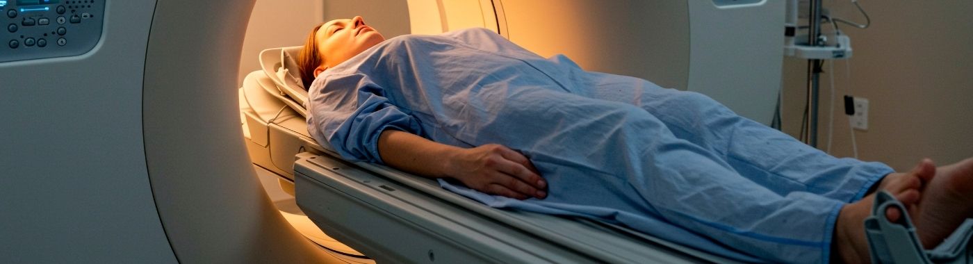
A cardiac PET scan is a nuclear imaging test that uses a short-lived radioactive tracer to map blood flow to the heart muscle, and sometimes its metabolism. Cardiac PET helps assess suspected coronary artery disease, myocardial viability, cardiac sarcoidosis and infection. Cardiac PET does not directly picture the coronary arteries themselves.
Panyawat konci
- PET shows function, not anatomy: the scan maps perfusion and metabolism of heart muscle, not the shape of the coronary arteries.
- Two different studies share the name: myocardial perfusion PET (blood flow) and FDG PET (viability, inflammation or infection) answer different questions.
- A normal scan is reassuring but not absolute: balanced three-vessel disease and non-obstructive plaque can be missed.
- Preparation differs sharply between study types: particularly the high-fat, no-carbohydrate diet before FDG studies for sarcoidosis.
- Hasilna rupa-rupa gumantung kana kaayaan klinis masing-masing and must be read by the doctor who requested the scan, alongside symptoms and other tests.
Sakolébat
| hal nu husus | kawincikan nepi ka bubuk leutikna |
|---|---|
| Nami lengkep | Cardiac positron emission tomography, usually done as PET/CT |
| Naon ukuranana | Myocardial blood flow, tracer uptake, glucose metabolism (depending on tracer) |
| Common tracers | Rubidium-82 or N-13 ammonia for perfusion; F-18 FDG for viability and inflammation |
| Invasiveness | Non-invasive apart from an intravenous cannula; stress studies use a pharmacological agent |
| radiasi | Yes, ionising radiation; dose depends on tracer and protocol |
| Typical time in department | About 1-3 hours in total; actual imaging usually 20-60 minutes |
| Availability in India | Limited to larger centres with a PET/CT scanner and cardiac-capable tracer supply |
Ogé katelah
- PET heart scan
- Myocardial perfusion PET / PET MPI
- Jantung PET/CT
- FDG PET heart scan, FDG viability scan
- Commonly said in India as "heart PET scan", "PET CT for heart", or grouped loosely with the "nuclear scan" or "thallium test" (the thallium/sestamibi study is actually SPECT, a different test)
What a cardiac PET scan is
A cardiac PET scan uses a positron-emitting tracer injected into a vein. The tracer travels to heart muscle in proportion to blood supply or metabolic activity. The scanner detects the signal and builds a three-dimensional map of the left ventricle. A low-dose CT is usually taken at the same time for attenuation correction, this CT is not a CT coronary angiogram and does not show the arteries in diagnostic detail unless a separate contrast study is specifically ordered.
Two broad study types exist, and they are not interchangeable:
- Perfusion PET (rest and stress): compares blood flow at rest with flow during pharmacological stress (commonly adenosine, regadenoson or dipyridamole). Many units also report myocardial blood flow in absolute terms and calculate myocardial flow reserve.
- FDG PET: uses a glucose analogue. With carbohydrate-loading protocols it shows whether poorly contracting muscle is still alive (viable). With a high-fat, very-low-carbohydrate preparation and fasting it is used instead to look for inflammation, as in suspected cardiac sarcoidosis, or for device and valve infection.
Why a cardiac PET scan is done
- Suspected obstructive coronary artery disease where a functional test is preferred, or where earlier tests were inconclusive.
- Myocardial viability before revascularisation in a weak heart muscle, to judge whether contraction may improve.
- Microvascular dysfunction, using absolute flow quantification, in people with angina and no obstructive disease on angiography.
- Sarkoidosis jantung: assessment of active inflammation and monitoring of response.
- Suspected device, graft or prosthetic valve infection, usually alongside echocardiography and blood cultures.
- Difficult imaging conditions: large body habitus, breast or diaphragmatic attenuation, where SPECT images are often degraded.
Saha anu kedah mertimbangkeun tés ieu
Cardiac PET is not a screening test for the general public or for people without symptoms. It is generally considered when a cardiologist has a specific question, such as:
- Chest pain or breathlessness with an intermediate likelihood of coronary disease, where an exercise ECG is not possible or was uninterpretable.
- Known coronary disease with recurrent symptoms, to judge which territory is ischaemic before considering stenting or bypass.
- Reduced ejection fraction with coronary disease, where viability influences whether revascularisation is offered.
- Angina with unobstructed coronary arteries, where microvascular disease is suspected.
- Unexplained heart block, ventricular arrhythmia or cardiomyopathy in a younger adult where sarcoidosis is being considered.
- People in whom a previous SPECT study was inconclusive because of attenuation artefact.
It is usually avoided or deferred in pregnancy, and used with careful justification in young people, because of radiation.
What a cardiac PET scan cannot detect or exclude
- Éta henteu ngabayangkeun arteri koroner. It cannot tell you the percentage narrowing of a specific artery. That requires CT coronary angiography or invasive angiography.
- It can under-call balanced ischaemia. If all three vessels are severely narrowed, relative perfusion may look uniform. Absolute flow quantification reduces but does not remove this problem.
- It does not detect non-obstructive plaque that has not yet limited flow, plaque that can still rupture and cause a heart attack.
- A normal perfusion PET does not exclude coronary artery disease, nor does it guarantee a future free of cardiac events.
- It is not a valve, aorta or pericardial test. Echocardiography and cardiac MRI answer those questions.
- FDG uptake is not specific. With inadequate dietary preparation, normal heart muscle takes up FDG and can mimic inflammation.
- A negative scan is "not identified", not "excluded". If your symptoms continue or worsen despite a normal result, go back to your doctor rather than assuming the heart has been cleared.
Ujian anu aya patalina tapi béda
These tests are often spoken of together but answer different questions. None is universally superior; the right first test depends on the clinical problem.
| ujian | Patarosan eta ngajawab | Watesan konci |
|---|---|---|
| Cardiac perfusion PET | Is blood flow to the heart muscle reduced, and by how much? | No coronary anatomy; radiation; limited availability |
| SPECT MPI (thallium/sestamibi "nuclear stress test") | Similar perfusion question, more widely available | Lower spatial resolution, more attenuation artefact, generally no absolute flow quantification |
| Angiografi koroner CT | Are the coronary arteries narrowed or plaque-laden? | Shows anatomy, not whether a narrowing limits flow; iodinated contrast; calcium can obscure |
| Angiografi koroner invasif | Anatomy with the option to treat in the same sitting | Invasive, small procedural risk |
| Resting 2D echocardiogram ("echo test") | Pump function, valves, chamber size, pericardium | Does not visualise coronary arteries; a normal echo does not exclude coronary disease |
| Ékokardiogram setrés | Wall motion change under stress; no radiation | Operator-dependent; image quality varies |
| Jantung MRI | Scar, fibrosis, myocarditis, infiltration, viability without radiation | Implants need MRI safety assessment; longer scan; breath-holds needed |
| FDG PET (inflammation protocol) | Is there active inflammation or infection in the heart? | Highly dependent on strict dietary preparation |
Cardiac PET and FDG viability PET are also distinct from each other, a request for one does not include the other.
Kumaha nyiapkeun
Preparation instructions are set by each imaging unit and depend on the tracer, the scanner and the clinical question. Follow the written instructions your unit gives you; those take precedence over anything on this page.
- puasa: most units ask for a period of fasting, commonly several hours, with plain water allowed. The exact duration is unit-specific.
- kafein: for stress perfusion studies using adenosine, regadenoson or dipyridamole, caffeine blocks the drug and can make the test uninterpretable. Units typically ask for a caffeine-free period beforehand, including tea, coffee, cola, chocolate, energy drinks and some headache tablets. Confirm the required duration with your unit.
- Tobacco and nicotine: usually asked to be avoided before the study.
- Special diet for FDG inflammation studies: a high-fat, very-low-carbohydrate diet for a day or more, then fasting, is used to suppress normal heart muscle glucose uptake. Skipping this often ruins the scan.
- obat: bring a full list, including AYUSH, herbal and gym supplements. Some cardiac medicines and theophylline-type drugs interact with stress agents. Do not start, stop or change any prescribed medicine yourself, ask the prescriber or the imaging unit.
- Clothing and metal: wear comfortable clothes without metal fastenings; you may be asked to change into a gown.
- Allergies and previous reactions: tell the team about any prior reaction to a tracer, contrast or stress agent.
Upami anjeun ngagaduhan diabetes anu nuju dirawat pikeun nurunkeun glukosa
Fasting and carbohydrate protocols can cause hypoglycaemia, and blood glucose strongly affects FDG images. Tell the unit in advance that you take insulin or other glucose-lowering medicines, ask for specific timing instructions, and carry a sugar source.
Upami anjeun ngalaman gagal jantung atanapi pangwatesan cairan
General advice to "drink plenty of water" afterwards is not appropriate for everyone. If you have heart failure, advanced chronic kidney disease or a prescribed fluid limit, follow the fluid allowance your treating doctor has given you.
If you have asthma, COPD or bradycardia
Adenosine-type stress agents can cause bronchospasm and heart block. Tell the team beforehand; an alternative agent or protocol may be chosen.
Upami anjeun hamil atanapi nyusuan
Tell the team before any tracer is given. Cardiac PET is generally avoided in pregnancy unless there is a compelling reason. If breastfeeding, ask for specific advice on expressing and interrupting feeds for the tracer used.
barudak
Cardiac PET in children is uncommon, done only in specialist centres, with weight-based tracer dosing and paediatric preparation and sedation protocols.
Naon anu kajantenan nalika scan
- Cék in: identity, pregnancy check where relevant, weight, blood glucose for FDG studies, and consent.
- Cannula and ECG: an intravenous line is placed; ECG leads and a blood pressure cuff are attached for stress studies.
- Tracer injection and uptake: the tracer is injected. Perfusion tracers image almost immediately; FDG needs an uptake period of roughly an hour of quiet rest.
- Rest imaging: you lie still on the scanner couch with your arms usually above your head.
- Stress phase (perfusion studies only): the stress drug is infused. Flushing, chest heaviness, breathlessness or a headache are common and usually settle within minutes. Tell the staff what you feel.
- Stress imaging: a second set of images is acquired.
- Salajengna: the cannula is removed once you are stable. Most people return to normal activity the same day unless told otherwise.
Sabaraha lami prosésna
- Rest-stress perfusion PET: often around 1 to 2 hours in the department, with imaging itself typically 20-40 minutes.
- FDG viability or inflammation PET: usually longer, commonly 2 to 3 hours, because of the uptake period.
- Dietary preparation for inflammation studies starts a day or more before the appointment.
Exact timings vary by unit, scanner and tracer supply.
Ngartos laporan anjeun
A cardiac PET report is descriptive and image-based rather than a list of numbers with a single normal range. Typical elements include:
- Perfusion pattern: uniform uptake suggests no regional flow limitation. A defect present on stress but not at rest suggests ischaemia; a defect present on both may suggest scar.
- Extent and severity: often expressed as a percentage of the left ventricle involved and mapped to vascular territories.
- Left ventricular function: ejection fraction at rest and, in some protocols, after stress.
- Myocardial blood flow and flow reserve: reported by some units; low global flow reserve can point to microvascular disease or diffuse disease.
- Viability (FDG studies): reduced perfusion with preserved FDG uptake, a flow-metabolism mismatch, suggests hibernating but living muscle.
- Inflammation (sarcoidosis protocol): focal or focal-on-diffuse FDG uptake, interpreted together with perfusion.
Grading and defect scoring involve reader judgement and vary between centres. A "mild" or "small" defect is not a diagnosis by itself. Cardiac PET results must be interpreted in clinical context by the cardiologist who requested the scan, together with your symptoms, ECG, echo and blood results. No treatment decision should be made from the report text alone.
Nalika hasilna tiasa nyasabkeun
Falsely suggesting disease (false positive or overcalled)
- Patient movement or misalignment between the PET and CT attenuation map, creating artefactual defects.
- Diaphragm, breast or arm attenuation not fully corrected.
- Left bundle branch block or a pacing rhythm, which can cause septal perfusion abnormalities without coronary disease.
- Inadequate carbohydrate restriction before an inflammation FDG study, so normal myocardium lights up.
- Recent heavy exercise or metabolic states altering FDG distribution.
Falsely reassuring (false negative or undercalled)
- Balanced three-vessel or left main disease producing apparently uniform relative perfusion.
- Caffeine or theophylline-type medicines blunting the stress agent, so true ischaemia is not provoked.
- Submaximal haemodynamic response to the stress drug.
- Small subendocardial ischaemia below the resolution of the technique.
- Extensive non-obstructive plaque, which does not limit flow and therefore does not show up.
Technical and biological factors
- High blood glucose or insulin at the time of FDG injection alters uptake.
- Very high body weight can degrade counts even on PET, though less than on SPECT.
- Arrhythmia during gated acquisition can make ejection fraction unreliable.
Résiko sareng kaamanan
radiasi
Cardiac PET uses ionising radiation. Doses depend heavily on the tracer and protocol, rubidium-82 and N-13 ammonia studies generally deliver lower doses than many SPECT protocols, while FDG studies with a diagnostic CT deliver more. The exposure is justified only when the result is likely to change management. Ask your team why the scan is needed and whether a radiation-free alternative such as stress echo or cardiac MRI would answer the same question.
Pharmacological stress agents
Adenosine, regadenoson and dipyridamole commonly cause transient flushing, chest tightness, breathlessness, nausea or headache. Less commonly they can cause bronchospasm, significant bradycardia or heart block, and rarely a serious cardiac event. These agents are given in a monitored setting with resuscitation facilities and reversal drugs available. Dobutamine protocols carry a different profile, including arrhythmia.
Tracer injection
Reactions to PET tracers are uncommon. Bruising or discomfort at the cannula site can occur. PET tracers are not iodinated contrast, so iodine allergy is not usually relevant unless a separate contrast CT is planned.
Practical discomfort
Lying still with arms raised can be uncomfortable, particularly with shoulder or back problems. Tell the radiographer, positioning support can often help.
Gendéra beureum
Emergency now: call 108 or go to the nearest emergency department
- Chest pain, pressure or heaviness lasting more than a few minutes, or spreading to the arm, jaw, neck or back.
- Severe breathlessness at rest, or sudden inability to speak in full sentences.
- Fainting, collapse, or a fast irregular heartbeat with dizziness.
- Sudden weakness of face, arm or leg, or slurred speech.
- Wheeze, facial or throat swelling, rash or lightheadedness after an injection.
Same day: contact your cardiologist or imaging unit
- Chest discomfort or palpitations that begin after you leave the department and then settle.
- Persistent headache, flushing or breathlessness hours after a stress agent.
- Increasing pain, swelling or redness at the cannula site.
- Angina that is occurring more often or at lower effort than before.
Janjian rutin
- Discussing your report and what it means for treatment.
- Mild bruising at the injection site.
- Stable symptoms that have not changed in pattern.
- Questions about whether and when the scan may need repeating.
kaayaan husus
reuneuh
Radiation is avoided in pregnancy wherever possible. Tell the team if you are or might be pregnant. Stress echocardiography or cardiac MRI without gadolinium are often preferred alternatives.
Breastfeeding
Advice depends on the tracer. Some require a short interruption of feeding; others do not. Ask the nuclear medicine team for tracer-specific instructions before the injection.
sawawa heubeul
Lying flat with arms raised, and the effects of stress agents, may be harder to tolerate. Frailty, arthritis and postural symptoms are worth flagging in advance.
Diabetes dina ubar anu nurunkeun glukosa
Blood glucose affects FDG images, and fasting affects glucose control. Ask the unit for individualised timing instructions for insulin and tablets rather than adjusting doses yourself.
panyakit ginjal kronis
PET tracers are not iodinated contrast and are generally used without the contrast-related kidney precautions. If a contrast-enhanced CT is being added, the kidney function will be reviewed separately. Fluid advice should follow your nephrologist's guidance.
gagalna jantung
Cardiac PET is often specifically used in heart failure to assess viability and cause. Post-scan fluid advice must respect any prescribed fluid restriction.
Asma sareng COPD
Adenosine and dipyridamole may be avoided in active bronchospastic disease. Bring your inhalers and tell the team about recent exacerbations.
Cardiac PET in the Indian context
- Availability is uneven. PET/CT scanners are concentrated in metros and large tertiary centres. Cardiac-specific tracers such as rubidium-82 generators or on-site cyclotron-produced N-13 ammonia are available in only a subset of these. Many centres offer FDG cardiac studies but not perfusion PET. Confirm before travelling.
- SPECT remains the more common nuclear perfusion test across the country, and is often referred to as the "thallium test" or "stress test with dye". It answers a similar question with lower resolution.
- Coronary disease in South Asians tends to present at a younger age and with metabolic risk at lower body mass index than in many Western populations, which affects how symptoms in young adults are taken seriously.
- Panyakit jantung rematik remains an important cause of cardiac symptoms in India. Valve disease is assessed with echocardiography, not PET, breathlessness in a young adult often warrants an echo first.
- Cardiac sarcoidosis and cardiac tuberculosis can both present with unexplained heart block or cardiomyopathy. FDG PET interpretation in India therefore needs careful clinical correlation.
- Aturan: facilities using radioisotopes operate under Atomic Energy Regulatory Board licensing, and clinical practice is governed by NMC professional conduct rules. Any online form or booking tool handling your health data falls under the Digital Personal Data Protection Act, 2023.
- basa: ask for your report explanation in the language you are most comfortable with; interpretation of an image-based report is easy to misread on your own.
Biaya sareng asuransi di India
Cardiac PET is among the more expensive cardiac imaging tests in India, typically costing substantially more than a stress echocardiogram or a SPECT perfusion scan. Prices differ widely between cities, between government and private centres, and between study types, so obtain a written quotation from the specific centre before booking. Any figure quoted online is indicative only and not a commitment.
Factors that affect the price:
- Tracer used, generator-produced rubidium-82 versus cyclotron-produced N-13 ammonia versus FDG, and how far the tracer must travel.
- Whether the study is rest-only, rest-stress, or viability with two tracers.
- Cost of the pharmacological stress agent.
- Whether a diagnostic contrast CT or calcium score is added.
- Scanner generation and whether absolute flow quantification software is used.
- City, hospital tier and whether the scan is done as an inpatient.
- Add-ons such as sedation, cardiologist supervision charges and repeat imaging.
asuransi: health insurers in India often cover cardiac PET when it is medically indicated and part of an admission or an approved day-care or diagnostic benefit, but outpatient diagnostics are excluded from many indemnity policies. Pre-authorisation with a clear clinical justification from your cardiologist improves the chance of approval. Check your specific policy wording and speak to the hospital's insurance desk before the scan.
Mitos sareng fakta
| carita | kanyataan |
|---|---|
| A PET scan shows my blocked arteries. | Cardiac PET shows blood flow to heart muscle, not the arteries themselves. Anatomy needs CT coronary angiography or invasive angiography. |
| A normal PET means my heart is fine for years. | A normal scan lowers but does not remove risk. Non-obstructive plaque can still cause events. Symptoms that persist need review. |
| PET is always better than a stress echo or SPECT. | Each test answers a different question with different trade-offs in radiation, availability and cost. The right test depends on the clinical problem. |
| The tracer will make me radioactive and dangerous to my family. | PET tracers decay quickly. Units give specific short-term advice about close contact with infants and pregnant women; follow that advice. |
| A PET scan is a good yearly heart check-up. | Cardiac PET is not a screening test. Using ionising radiation without a clinical question is not justified. |
| Skipping the special diet before an FDG scan is no big deal. | Poor dietary preparation is the commonest reason an FDG inflammation scan is uninterpretable and has to be repeated. |
Remen ditanyakeun
Is a cardiac PET scan better than an angiography?
Neither is better; they answer different questions. Cardiac PET shows whether blood flow to the heart muscle is reduced. Angiography shows the anatomy of the coronary arteries and allows treatment in the same sitting. Cardiologists often use a functional test like PET to decide whether angiography is needed at all.
Why was I told to avoid tea and coffee before the scan?
Caffeine blocks adenosine-type stress agents at the receptor level. If caffeine is present, the drug may not increase blood flow enough, and genuine ischaemia can be missed. This is why units ask for a caffeine-free period. The required duration differs between units, so follow your written instruction.
Will the stress injection cause a heart attack?
Serious events are uncommon. Flushing, chest heaviness, breathlessness and headache are common and usually pass within minutes. The test is performed with continuous monitoring and resuscitation facilities. Tell the staff immediately about any symptom rather than waiting for the scan to finish.
How much radiation does a cardiac PET involve?
The dose depends on tracer and protocol, and is not a single fixed number. Some PET perfusion protocols involve less radiation than conventional SPECT nuclear scans, while FDG studies with a diagnostic CT involve more. Ask your nuclear medicine team for the estimated dose for your specific protocol.
Can a cardiac PET scan be repeated?
Yes, when there is a clear clinical reason, such as monitoring treatment for cardiac sarcoidosis. Repeat imaging is not scheduled routinely, because each scan adds radiation exposure. The interval is decided by your cardiologist based on the question being asked.
What does "flow-metabolism mismatch" mean on my report?
The phrase describes an area with reduced blood flow but preserved glucose uptake on FDG imaging. It suggests heart muscle that is underperfused but still alive, sometimes called hibernating myocardium. Such areas may improve after revascularisation, though outcomes vary depending on individual clinical circumstances.
Do I need someone to accompany me?
It is sensible. You may feel washed out after a stress agent, the appointment can run to several hours, and having someone to drive you home is safer. Some units require an accompanying adult for sedation or for older patients, check when booking.
Can I eat and drink normally afterwards?
Most people can resume a normal diet immediately unless told otherwise. Fluids help the tracer clear, but if you have heart failure, advanced kidney disease or a prescribed fluid restriction, stay within the allowance your treating doctor has set rather than drinking extra.
My scan was normal but I still get chest pain. What now?
Return to your doctor. A normal perfusion scan means no significant flow-limiting disease was identified; it does not exclude coronary disease, microvascular angina, or non-cardiac causes such as reflux, musculoskeletal pain or anxiety. Persistent or worsening symptoms deserve reassessment rather than reassurance based on an old result.
sumber
- American Society of Nuclear Cardiology and Society of Nuclear Medicine and Molecular Imaging, joint guidance and information statements on cardiac PET myocardial perfusion and metabolism imaging.
- Heart Rhythm Society expert consensus statement on the recognition and management of arrhythmias associated with cardiac sarcoidosis.
- European Society of Cardiology guidelines on the management of chronic coronary syndromes.
- American College of Cardiology / American Heart Association guideline on the evaluation and diagnosis of chest pain.
- Atomic Energy Regulatory Board (India), regulatory requirements for medical use of radioisotopes.
- Digital Personal Data Protection Act, 2023 (India).










 Rumah Sakit Pangsaéna di Chennai


